Zhu's Scalp Acupuncture: Navigating Cranial Treatment Zones, Daoyin Somatokinetic Facilitation, and Precision Acupressure Protocols
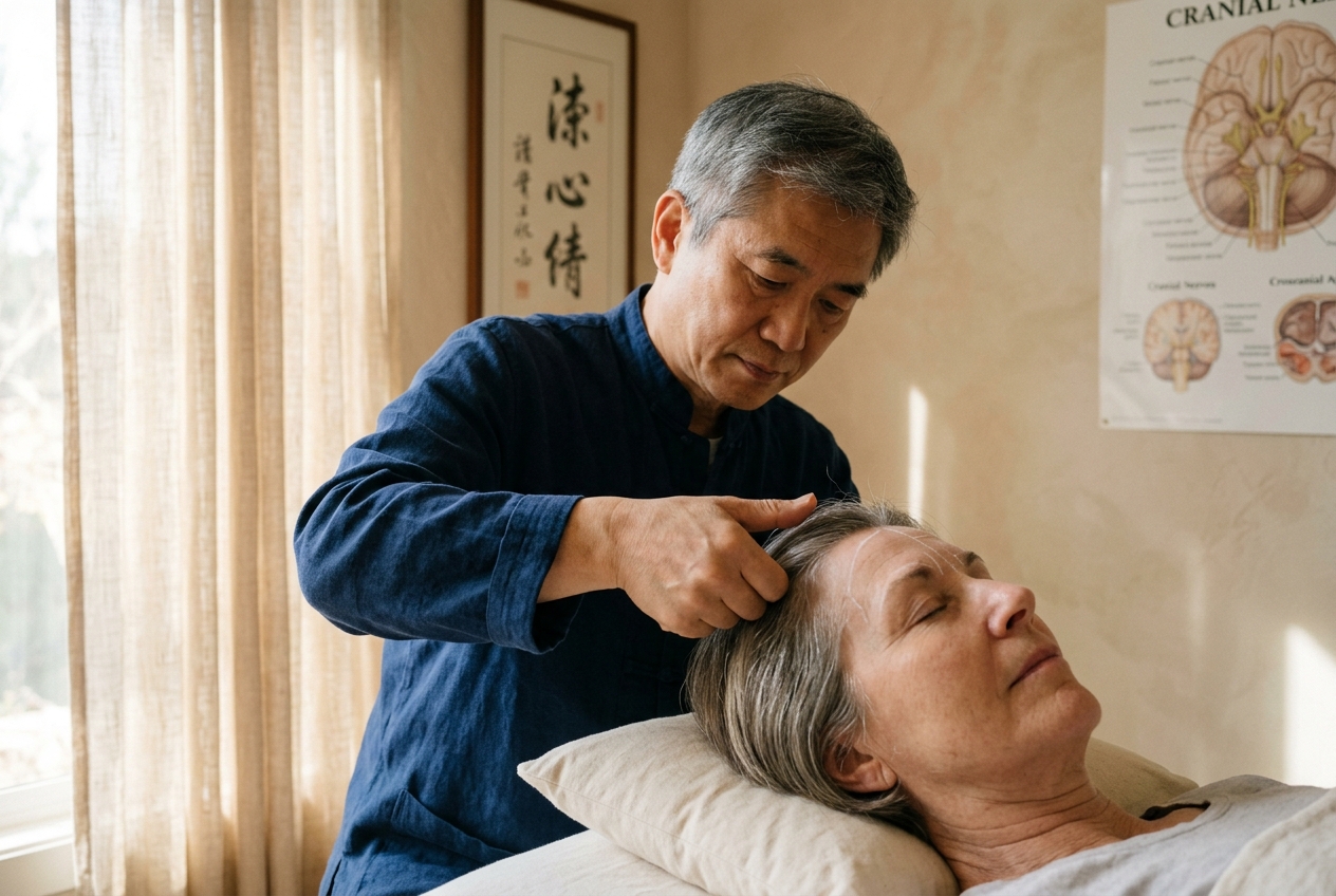
Developed over five decades of clinical inquiry by Dr. Ming Qing Zhu, this specialized neuro-acupuncture discipline synthesizes the meridian pathways of classical Chinese medicine with the somatotopic maps of contemporary neuroanatomy. Unlike conventional body acupuncture or passive cranial modalities, Zhu’s method pairs precise cranial stimulation with immediate, active somatic kinetic movement—a principle known as Daoyin. Whether administered through filiform needles or high-precision non-invasive manual acupressure, Zhu’s Scalp Acupuncture provides clinicians and patients with a targeted protocol to stimulate collateral circulation, promote neuroplastic remodeling, and restore lost functional capacity.
1. Foundations & Lineage: The Synthesis of Ling Shu Channel Dynamics and Cortical Localization
The historical trajectory of scalp acupuncture is characterized by a continuous dialogue between classical meridian energetics and empirical cortical mapping. In classical texts such as the Huangdi Neijing Ling Shu (circa 100 BCE), the cranium is identified as the "Sea of Marrow" (Sui Hai), the biological nexus where all primary Yang channels converge to nourish the brain and govern sensory perception. The Ling Shu (Chapter 33) explicitly notes that when the Sea of Marrow is replete, the body exhibits vitality, sharp sensory acuity, and physical agility; when deficient, it manifests as dizziness, tinnitus, gait instability, and motor lethargy.
During the 1970s, the emergence of modern scalp acupuncture in China gave rise to distinct methodologies. Dr. Jiao Shunfa developed a system based strictly on anatomical surface projections of the underlying cerebral cortex, delineating rigid motor and sensory strips derived from Wilder Penfield’s motor homunculus. Concurrently, in Japan, Dr. Toshikatsu Yamamoto established Yamamoto New Scalp Acupuncture (YNSA), a distinct microsystem utilizing localized diagnostic and therapeutic points across the frontal and temporal scalp regions.
Dr. Ming Qing Zhu departed fundamentally from these models. While acknowledging Penfield’s cerebral localization, Dr. Zhu recognized that functional neurological recovery requires a dynamic, fluid, and continuous approach. Rather than relying on discrete isolated points or rigid cerebral projection lines, Zhu established continuous functional cranial zones. These zones trace the natural fascial planes and meridian trajectories of the scalp, notably along the Governing Vessel (Du Mai), Bladder (Taiyang), and Gallbladder (Shaoyang) meridians.
Crucially, while Jiao’s and Yamamoto’s protocols traditionally require the patient to remain motionless during treatment, Dr. Zhu instituted the mandatory integration of simultaneous active movement (Daoyin). In Zhu’s paradigm, cranial stimulation does not merely sedate or tonify an inert system; it opens a transient neurovascular window of opportunity during which the patient must actively engage the paretic or dysfunctional limb to drive functional cortical reorganization.
2. Neurofascial & Bio-Kinetic Mechanisms: Galea Aponeurotica, Pericranial Perfusion, and Neurovascular Coupling
To understand how mechanical pressure or needle stimulation on the scalp alters deep cerebral function, one must examine the micro-anatomical architecture of the cranial vault. The scalp comprises five distinct layers, remembered through the mnemonic SCALP: Skin, Connective tissue (dense subcutaneous layer), Aponeurosis (galea aponeurotica), Loose subaponeurotic connective tissue, and Pericranium (periosteum).
The Galea Aponeurotica as a Piezoelectric Transmission Network
The galea aponeurotica is a broad, dense sheet of collagenous tendon that connects the frontalis muscle anteriorly to the occipitalis muscle posteriorly, integrating laterally with the temporoparietal fascia. When manual pressure, directional shear, or horizontal needle manipulation is applied tangentially across the subgaleal plane, several biological cascades are triggered:
- Piezoelectric Transduction: Mechanical shear deformation of the dense collagen matrix in the galea generates micro-electrical surface charges. These electrical potentials stimulate underlying unmyelinated C-fibers and myelinated A-beta mechanoreceptors embedded within the pericranial fascia.
- Subgaleal Microcirculation & Emissary Communication: Compressive force along the loose subaponeurotic plane alters hemodynamic pressure within the pericranial vascular arcade. The cranial vault contains valveless emissary veins that traverse the diploic space of the skull bones, directly connecting extracranial scalp veins to the intracranial dural venous sinuses. Mechanical deformation of these tissues influences local pericranial drainage and modulates dural venous compliance.
Trigeminocervical Sensory Loops and Autonomic Modulation
The anterior two-thirds of the scalp are innervated by the ophthalmic ($V_1$), maxillary ($V_2$), and mandibular ($V_3$) divisions of the trigeminal nerve, while the posterior third is innervated by the greater and lesser occipital nerves originating from the cervical plexus ($C_2, C_3$).
Stimulation across Zhu’s cranial zones converges upon the trigeminocervical complex in the upper cervical spinal cord and brainstem. This sensory influx projects directly into the reticular activating system, the solitary tract nucleus (nucleus tractus solitarii), and the thalamus. This pathway drives an immediate sympatholytic effect, downregulating systemic sympathetic hyperarousal while upregulating parasympathetic vagal tone. Research published in Frontiers in Neuroscience demonstrates that targeted sensory stimulation of cranial trigeminal branches enhances cerebral autoregulation and modulates the diameter of middle cerebral arteries via the trigeminovascular reflex.
Neurovascular Coupling and Penumbral Rescue
According to the principle of neurovascular coupling, local neural activity is directly coupled to cerebral blood flow via endothelial nitric oxide synthase (eNOS) activation and astroglial foot-process signaling. In ischemic stroke, the central necrotic core is surrounded by an ischemic penumbra—a zone of electrically silent but metabolically viable brain tissue.
By stimulating the precise somatotopic cranial zone corresponding to the injured cortical area, subgaleal mechanoreceptor firing drives focal functional hyperemia, promoting oxygenation and glucose delivery to these vulnerable penumbral neurons, thereby preventing secondary apoptotic cascade progression.
3. The Daoyin Dynamic Movement Principle: Neuromuscular Re-Education and Cortical Plasticity
The definitive clinical hallmark of Zhu’s Scalp Acupuncture is the simultaneous application of Daoyin (guided somatic intentionality and dynamic movement). In conventional acupuncture, patients are typically instructed to remain motionless in a supine or prone posture to prevent needle deformation. Dr. Zhu overturned this passive convention, demonstrating that the therapeutic efficacy of scalp stimulation is magnified when delivered synchronously with active motor-sensory execution.
The Neuroplastic Mechanism of Synchronous Stimulation
Modern neurorehabilitation confirms that damaged central neural pathways undergo functional reorganization through neuroplasticity, governed largely by Hebbian learning principles: "neurons that fire together, wire together."
When a patient suffers an upper motor neuron lesion (such as in an ischemic or hemorrhagic stroke), descending corticospinal drive is interrupted. The cortical representation of the affected limb shrinks rapidly as a consequence of "learned non-use."
When manual acupressure or filiform needling is applied to Zhu's Dingnie (Motor-Sensory) Zone: 1. The cranial stimulation acts as a neuromodulatory primer, depolarizing local neuronal assemblies within the sensorimotor cortex and lowering the threshold required for synaptic firing. 2. If the patient simultaneously attempts to flex the paretic elbow, dorsiflex the paralyzed ankle, or articulate phonemes during this excitation window, the ascending proprioceptive and kinesthetic feedback loops from the periphery arrive at the cerebral cortex at the precise instant the cortical zone is being activated. 3. This synchronous dual-input signal establishes immediate sensorimotor loop coherence, promoting long-term potentiation (LTP) across latent synaptic connections, upregulating brain-derived neurotrophic factor (BDNF), and stimulating perilesional cortical areas to assume the functional duties of the infarcted tissue.
Structured Daoyin Protocol During Cranial Stimulation
A standard clinical Daoyin sequence consists of three integrated components:
- Rhythmic Diaphragmatic Respiration: The patient is instructed to inhale deeply through the nose, expanding the lower abdomen, and exhale slowly through slightly parted lips. This breathing resets autonomic tone and synchronizes cerebral blood flow oscillations.
- Active-Assisted Kinetic Engagement: The clinician or caregiver assists the patient in executing active range-of-motion (AROM) drills through the paretic joint. If the patient possesses zero trace muscle contraction (Grade 0 on the Oxford Muscle Scale), mental motor imagery (visualizing the limb moving with absolute clarity) is paired with passive therapist-guided motion.
- Progressive Resistance and Functional Tasks: As trace contractions (Grade 1–2) return, the patient executes functional motor patterns—such as grasping a cup, weight-bearing on the paretic heel, or phonating targeted consonant-vowel combinations—while continuous manual pressure or needle stimulation is maintained on the scalp.
4. Topographical Cartography of Core Zhu Treatment Zones
Zhu’s scalp topography is arranged into distinct functional zones corresponding to specific clinical territories of the human body and central nervous system. Precise localization relies on palpable cranial landmarks: the glabella, the external occipital protuberance (inion), the apex of the auricle, and the cranial sutures (coronal, sagittal, and lambdoid).
A. Dingzhong Zone (Vertex Center Zone / 顶中区)
- Anatomical Boundaries: Extends along the sagittal midline of the cranium, spanning a 1.5-cun linear path between Baihui (GV20) and Qianding (GV21).
- Underlying Neuroanatomy: Directly overlies the longitudinal cerebral fissure, the superior sagittal sinus, and the paracentral lobules of the primary motor and primary somatosensory cortices.
- Somatotopic Correspondences: Corresponds somatotopically to the lumbosacral spine, pelvis, perineum, lower limbs, urinary bladder, and genitourinary organs.
- Clinical Indications: Essential for treating lower extremity paraparesis, acute lumbar radiculopathy, post-stroke urinary incontinence, neurogenic bladder dysfunction, and pelvic floor spasm.
B. Dingnie Zone (Vertex-Temporal Zone / 顶颞区)
- Anatomical Boundaries: A diagonal linear band extending from Baihui (GV20) obliquely anteroinferiorly toward Xuanli (GB6) along the temporal border. This zone is functionally divided into three equal segments:
- Upper One-Third: Originates at GV20 and extends one-third of the distance toward GB6.
- Middle One-Third: The central third of the diagonal trajectory.
- Lower One-Third: The inferior third terminating near GB6 on the temporal scalp.
- Underlying Neuroanatomy: Precisely mirrors the orientation of the precentral gyrus (primary motor cortex, Brodmann Area 4) and postcentral gyrus (primary somatosensory cortex, Brodmann Areas 1, 2, and 3).
- Somatotopic Division & Functional Mapping:
- Upper Third: Governs the contralateral hip, knee, ankle, foot, toes, and lower lumbar region.
- Middle Third: Governs the contralateral shoulder, elbow, wrist, hand, and fingers.
- Lower Third: Governs the contralateral facial musculature, tongue, pharynx, larynx, and Broca’s motor speech area.
- Clinical Indications: The primary therapeutic zone for post-stroke hemiplegia, upper extremity spasticity, flaccid hand paralysis, central facial palsy, dysphagia, and expressive (Broca’s) aphasia.
C. Dingfang Zone (Vertex-Lateral Zone / 顶旁区)
- Anatomical Boundaries: Bilateral linear strips positioned 0.5 to 1.0 cun lateral to the Dingzhong Zone on the parietal bones, running parallel to the sagittal midline.
- Underlying Neuroanatomy: Overlies the parietal association cortices and collateral vascular projections of the middle and anterior cerebral artery watershed zones.
- Somatotopic Correspondences: Corresponds to the thoracic cavity, upper burner (Shang Jiao), lungs, heart, and mediastinal viscera.
- Clinical Indications: Cardiorespiratory dysregulation, post-stroke chest tightness, neurogenic hyperventilation, chronic autonomic dysreflexia, and functional thoracic pain.
D. Ezhong Zone & Efang Zones (Forehead Center and Lateral Zones / 额中区与额旁区)
- Anatomical Boundaries:
- Ezhong Zone: A 1-cun vertical band extending superiorly along the anterior midline from the hairline (Shenting, GV24) upward.
- Efang 1 Zone: Bilateral vertical lines originating at the anterior hairline directly superior to the inner canthi of the eyes.
- Efang 2 Zone: Bilateral vertical lines originating at the anterior hairline directly superior to the outer canthi of the eyes.
- Underlying Neuroanatomy: Overlies the anterior prefrontal cortex (Brodmann Areas 9, 10, and 46) and the frontal eye fields (Brodmann Area 8).
- Functional Mapping: Governs executive cognitive functioning, emotional homeostasis, working memory, attention, and sensory orifice reception (ophthalmic and nasal tracts).
- Clinical Indications: Post-stroke depression, executive dysfunction, post-concussive syndrome, generalized anxiety disorder, central insomnia, and sensory integration deficits.
E. Zhenxia Zone (Sub-Occipital Zone / 枕下区)
- Anatomical Boundaries: Located on the posterior occiput, extending along the midline from the external occipital protuberance (Inion / Naohu, GV17) inferiorly to the superior border of the foramen magnum (Fengfu, GV16).
- Underlying Neuroanatomy: Directly overlies the cerebellar vermis, cerebellar hemispheres, and the dorsal brainstem (pons and medulla oblongata).
- Functional Mapping: Central equilibrium, vestibular coordination, muscle tone regulation, reciprocal antagonist inhibition, and gait stability.
- Clinical Indications: Cerebellar ataxia, post-stroke intentional tremor, vertigo, wall-walking gait, dysmetria, and nystagmus.
Zone Comparison & Clinical Mapping Guide
| Treatment Zone | Palpable Cranial Landmarks | Neuroanatomical Correlate | Primary Somatosensory Domain | Key Clinical Target |
|---|---|---|---|---|
| Dingzhong (Vertex Center) | Midline sagittal suture between GV20 and GV21 | Paracentral lobule; longitudinal fissure | Lumbosacral spine, pelvic floor, lower extremities | Paraplegia, urinary incontinence, sciatica |
| Dingnie (Upper 1/3) | Superior third of diagonal line from GV20 to GB6 | Superior pre/postcentral gyri | Contralateral hip, knee, ankle, and toes | Post-stroke foot drop, lower limb paresis |
| Dingnie (Middle 1/3) | Central third of diagonal line from GV20 to GB6 | Mid-lateral pre/postcentral gyri | Contralateral shoulder, arm, hand, fingers | Loss of fine motor grip, arm spasticity |
| Dingnie (Lower 1/3) | Inferior third of diagonal line from GV20 to GB6 | Inferior precentral gyrus, Broca’s area | Craniofacial muscles, tongue, vocal cords | Expressive aphasia, dysphagia, facial droop |
| Dingfang (Vertex Lateral) | 0.5–1 cun lateral to Dingzhong, parallel to midline | Parieto-occipital association cortex | Thoracic viscera, cardiopulmonary systems | Autonomic dysregulation, chest constriction |
| Ezhong / Efang (Forehead) | Hairline above glabella, inner canthi, & outer canthi | Anterior prefrontal cortex (Brodmann 9/10/46) | Executive function, mood, sensory orifices | Post-concussive fog, depression, anxiety |
| Zhenxia (Sub-Occipital) | Occipital midline from Inion (GV17) to GV16 | Cerebellar vermis, dorsal brainstem | Balance, coordination, equilibrium | Cerebellar gait ataxia, tremors, vertigo |
5. Precision Non-Invasive Acupressure Protocols
While Zhu’s Scalp Acupuncture is renowned for its filiform needle technique utilizing specialized short, horizontal sub-aponeurotic insertions, the same neurofascial and somatotopical mechanisms can be mobilized non-invasively through manual subgaleal acupressure.
This manual approach is valuable in clinical settings where needling is contraindicated (such as acute post-thrombolytic states or needle phobia) and serves as a vital self-care and caregiver intervention.
Palpation Rules: Identifying the Active Cranial Locus (Ah-Shi Nodes)
Before applying pressure, the practitioner must systematically palpate the designated cranial zone. A dormant zone will feel smooth, pliable, and non-reactive. In contrast, a zone linked to active neuropathology or acute pain exhibits distinct tactile tissue alterations: * Subgaleal Taut Bands: String-like, hypertonic collagenous ridges running parallel or transverse to the suture lines. * Spongy Edematous Pockets: Focal areas of fluid retention in the loose subaponeurotic layer. * Active Tender Loci (Ah-Shi points): Discrete points within the zone that elicit a sharp, distinct ache or radiating sensation upon moderate pressure. These points represent epicenters of neurogenic inflammation and pericranial receptor concentration.
Three Core Manual Manipulation Techniques
1. Linear Gliding Vector Compression (Tui Fa)
- Hand Position: Place the broad, fleshy pad of the thumb (or reinforced index and middle finger pads) directly at the origin of the target scalp zone.
- Vector and Depth: Depress the skin firmly until engaging the resistance of the galea aponeurotica. Maintain this depth without sliding over the epidermal surface; instead, glide the skin and underlying subcutaneous fascia across the periosteum in a continuous, unidirectional linear vector.
- Cadence: Apply firm, continuous pressure along the 1 to 1.5 cun length of the zone, moving at a controlled speed of 1 centimeter per 3–5 seconds. Repeat this gliding pass 10 to 15 times.
2. Targeted Ischemic Sustained Compression (An Fa)
- Execution: Upon isolating a distinct, tender Ah-Shi nodule within the zone, apply perpendicular sustained pressure using the tip or radial edge of the thumb.
- Intensity: Calibrate pressure to a firm, therapeutic intensity (a deep, localized dull ache, rated approximately 4 to 6 on a 10-point discomfort scale, avoiding sharp distress).
- Duration: Hold continuously for 60 to 90 seconds while instructing the patient to direct conscious awareness and diaphragmatic breath into the area.
3. Cross-Fiber Rhythmic Galeal Mobilization (Rou Fa)
- Execution: Anchor the thumb pad over the active nodule or suture intersection. Execute tiny, rhythmic circular and transverse shearing oscillations (1–2 mm amplitudes) at a rate of 2 to 3 Hz.
- Objective: Mobilizes the epicranial aponeurosis, releases subgaleal micro-adhesions, and stimulates mechanoreceptors to downregulate spinal reflex hypersensitivity.
6. Evidence-Based Clinical Applications & Stroke Rehabilitation Protocols
Clinical trials indexed on PubMed / NCBI have documented the efficacy of scalp acupuncture in treating central nervous system disorders, particularly as an adjunctive modality in post-stroke motor recovery, central neuropathic pain syndromes, and traumatic brain injury rehabilitation.
Protocol 1: Post-Stroke Hemiplegia & Upper Limb Flaccidity
- Primary Zones: Contralateral Dingnie Zone (Middle One-Third); Bilateral Dingzhong Zone.
- Secondary Zone: Ipsilateral Zhenxia Zone (for kinetic coordination).
- Manual Technique:
- Apply 2 minutes of linear gliding compression along the contralateral Middle Dingnie Zone.
- Identify the most reactive Ah-Shi point along the band; apply sustained ischemic compression.
- Synchronous Daoyin Drill:
- While pressure is maintained on the Middle Dingnie Zone, the patient sits upright and attempts active wrist extension, finger abduction, and elbow flexion.
- If no active movement exists, the therapist passively moves the patient's wrist into full extension while the patient concentrates on the kinesthetic sensation.
- Perform 3 sets of 10 movement cycles during continuous stimulation.
Protocol 2: Post-Stroke Expressive (Broca’s) Aphasia & Dysphagia
- Primary Zone: Contralateral (Dominant Hemisphere, typically left) Dingnie Zone (Lower One-Third).
- Secondary Zone: Bilateral Ezhong Zone.
- Manual Technique:
- Mobilize the lower third of the Dingnie zone (near the anterior-superior margin of the ear) using rhythmic cross-fiber friction for 90 seconds.
- Transition into linear gliding along the Ezhong Zone on the forehead for 60 seconds.
- Synchronous Daoyin Drill:
- While applying sustained pressure to the Lower Dingnie Zone, instruct the patient to articulate simple vowel sounds ("Ah", "Oh", "Ee"), count sequentially from 1 to 10, or practice swallowing small, measured sips of warm water.
- The direct activation of Broca’s projection zone during speech attempts recruits perilesional premotor networks to rebuild phonetic motor pathways.
Protocol 3: Central Spasticity Reduction Protocol
- Primary Zones: Bilateral Dingzhong Zone and Bilateral Zhenxia Zone.
- Mechanism: Spasticity represents a loss of upper motor neuron inhibitory control, leading to hyperactive stretch reflexes. Stimulating the midline vertex (Dingzhong) and sub-occipital cerebellar zones (Zhenxia) stimulates central inhibitory pathways and normalizes gamma motor neuron excitability.
- Procedure:
- Apply rhythmic thumb-kneading to the Dingzhong zone for 2 minutes.
- Shift to the Zhenxia zone below the inion, applying deep, slow circular friction for 2 minutes.
- Concurrently, perform slow, sustained, passive elongation of the spastic muscle groups (e.g., stretching the hypertonic biceps or gastrocnemius), holding each stretch at end-range for 30 seconds.
7. Clinical Safety, Contraindications, and Hemodynamic Precautions
While non-invasive cranial acupressure eliminates the risks of bleeding, subaponeurotic hematoma, or pneumocephalus associated with invasive filiform needling, rigorous clinical safety guidelines must be maintained:
Absolute Contraindications
- Unhealed Surgical Craniotomy or Craniectomy Sites: Never apply manual pressure or needle stimulation over a cranial bone defect, burr hole, or unhealed surgical flap. Maintain a minimum margin of 3 centimeters from any surgical margin.
- Acute Traumatic Skull Fracture: Any suspicion of open, linear, or depressed cranial fracture precludes scalp compression due to the risk of bone displacement or dural laceration.
- Severe Localized Scalp Infections: Cellulitis, fungal ulcerations, or active carbuncles on the scalp prohibit local manipulation to prevent intracranial spread via emissary veins.
Hemodynamic and Clinical Precautions
- Labile Hypertension: In patients with severe, uncontrolled hypertension (systolic BP > 180 mmHg or diastolic BP > 110 mmHg), avoid aggressive sub-occipital stimulation (Zhenxia Zone), which can transiently alter intracranial perfusion pressures. Begin with gentle, sedative gliding on the anterior forehead (Ezhong Zone) to induce autonomic down-regulation before addressing motor zones.
- Acute Ischemic Stroke Timing: According to clinical stroke guidelines from the National Institute of Neurological Disorders and Stroke (NINDS), acute ischemic stroke patients receiving intravenous tissue plasminogen activator (tPA) require gentle, non-invasive handling during the initial 24 hours post-infusion. Gentle manual acupressure and Daoyin can be initiated once hemodynamic and neurological stability is confirmed by the medical team.
- Pressure Titration: Pressure should be firm, rhythmic, and therapeutic. Avoid violent, abrupt percussive forces on the cranial vault. If the patient reports dizziness, lightheadedness, nausea, or localized sharp lancinating pain, discontinue stimulation immediately and place the patient in a comfortable supine position.
8. Self-Care Translational Protocol: The 4-Minute Daily Neurovascular Reset
For individuals dealing with cognitive fatigue, tension headaches, or chronic neck-shoulder stiffness from prolonged screen exposure, this practical sequence translates the principles of Zhu's Scalp Acupuncture into an effective daily self-care routine.
Step 1: Cognitive Decompression via the Ezhong Zone (Minute 1)
- Location: Place the pads of your index and middle fingers together at the center of your forehead, 1/2 inch above the midpoint of your eyebrows.
- Technique: Apply firm, upward gliding pressure straight up to the natural hairline.
- Action: Inhale deeply for a count of 4 through the nose, feeling the upward stretch of the forehead tissue; exhale slowly for a count of 6. Perform 10 continuous upward passes.
- Benefit: Alleviates frontal sinus congestion, eyestrain, and mental fatigue by stimulating prefrontal sensory pathways.
Step 2: Somatosensory Reset via the Dingnie Zone (Minutes 2 & 3)
- Location: Locate the vertex of your head (Baihui, the center point on top of the head aligned with the tips of the ears). Place your thumb pad 1 inch anterior to this point, angled diagonally down toward the top edge of your ear.
- Technique: Apply deep, circular friction using your thumb pad along this diagonal line, pausing at any tender, tight spots.
- Synchronous Daoyin Movement: While holding firm pressure on this tender spot, slowly roll your shoulders in broad circles and gently turn your head from side to side.
- Benefit: Inhibits muscle guarding in the upper trapezius, levator scapulae, and cervical spine via reciprocal sensory feedback.
Step 3: Cerebellar Equilibrium Reset via the Zhenxia Zone (Minute 4)
- Location: Slide your thumbs to the base of your skull on either side of the midline, resting in the hollows beneath the occipital ridge (Inion area).
- Technique: Hook your thumbs upward and inward under the bone ledge. Apply moderate, sustained pressure while maintaining a straight cervical spine.
- Action: Take three deep diaphragmatic breaths, gently tucking your chin toward your chest on the exhalation to gently stretch the sub-occipital muscle insertions.
- Benefit: Improves venous drainage through the sub-occipital arcade, reduces cervicogenic tension, and clears mental fog.
9. Conclusion: The Future of Cranial Neuromodulation
Zhu’s Scalp Acupuncture represents a remarkable convergence of classical empirical medicine and functional neurobiology. By viewing the cranium as a dynamic, responsive neurofascial switchboard rather than a static bone container, Dr. Ming Qing Zhu provided a transformative framework for central nervous system rehabilitation.
The integration of continuous topographical zones, subgaleal mechanoreceptor stimulation, and synchronous Daoyin kinetic re-education bridges the gap between passive therapeutic intervention and active neuroplastic restoration. As clinical research continues to illuminate the complex pathways of neurovascular coupling and the piezoelectric mechanics of the human fascia, Zhu’s methodology stands as an enduring, scientifically coherent blueprint for waking the brain’s dormant healing capacity.
Authoritative References & Further Reading
- World Health Organization: Standard Acupuncture Nomenclature — Global standards for cranial topography and meridian point localization.
- National Center for Biotechnology Information (NCBI / PubMed) — Clinical trials and systematic reviews on scalp acupuncture in post-stroke rehabilitation and motor recovery.
- National Institute of Neurological Disorders and Stroke (NINDS) — Research benchmarks for neuroplasticity, stroke recovery, and upper motor neuron lesions.
- Brain: A Journal of Neurology (Oxford Academic) — Foundational studies on neurovascular coupling, cortical mapping, and penumbral physiology.
- Galea Aponeurotica and Epicranial Fascial Biomechanics — Morphological and functional anatomy of the cranial neurofascial planes.