Yamamoto New Scalp Acupuncture: Navigating Basic and Ypsilon Somatotopic Topography, Cervical Diagnostic Zones, and Precision Acupressure Protocols
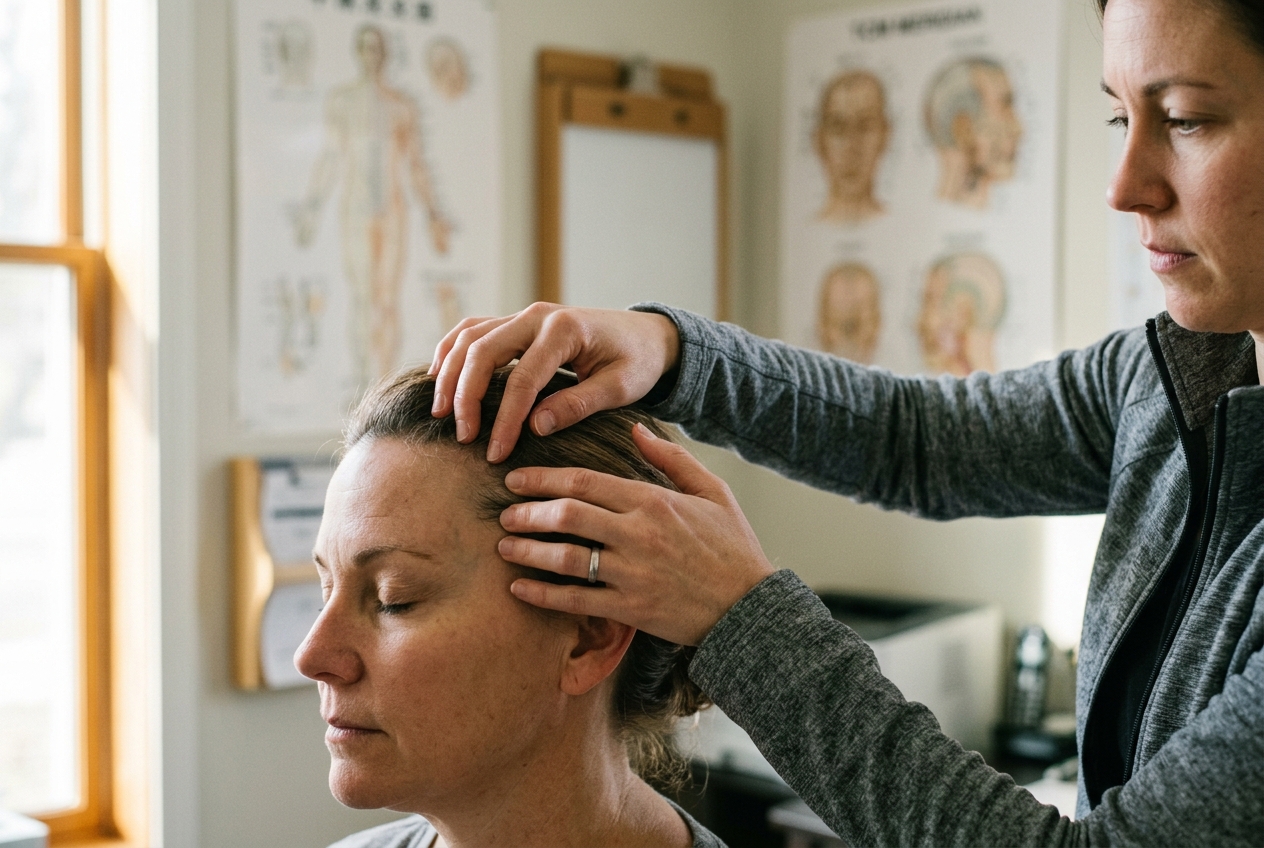
1. Opening — A Problem This Solves Today: The Neuro-Mechanical Crisis of the Modern Axial Skeleton and Central Paresis
If you wake with a grinding, intractable tightness at the craniocervical junction—an unyielding spasm where the base of the skull meets the cervical vertebrae that radiates downward into the trapezius and deadens the fingertips after hours of static digital posture—you are experiencing a profound breakdown in neuromotor communication. In more severe clinical presentations, such as the spastic hemiparesis that follows an ischemic cerebrovascular accident or the chronic radiculopathy of cervical spondylotic bi-syndrome, the central nervous system becomes locked in an aberrant loop of hypertonicity, proprioceptive distortion, and uncalibrated nociceptive signaling.
Conventional manual therapies often target these afflictions locally at the site of peripheral expression, kneading exhausted muscular bellies or tractioning inflamed facet joints. Yet, the neuromuscular command center remains refractory. Classical East Asian medicine, documented extensively through the World Health Organization's Standard Acupuncture Nomenclature, has long recognized that the periphery is merely an antenna; the master controls reside within specialized micro-somatotopic projections across the cranium.
+-----------------------------------------------------------------------------------+
| CLINICAL PROOF & THERAPEUTIC HORIZON                                              |
| Yamamoto New Scalp Acupuncture (YNSA) provides an immediate, empirically          |
| verifiable feedback loop. By stimulating precise 1–2 mm micro-somatotopic loci on  |
| the galea aponeurotica, practitioners can instantly extinguish myofascial         |
| indurations and tenderness in the cervical triangles (Neck Diagnosis), bypassing  |
| peripheral spinal bottlenecks and directly modulating thalamocortical pathways.   |
+-----------------------------------------------------------------------------------+
Developed in the early 1970s by the Japanese anesthesiologist and obstetrician Dr. Toshikatsu Yamamoto, Yamamoto New Scalp Acupuncture (YNSA) represents a radical departure from traditional Chinese channel systems while honoring the fundamental doctrine of Zang-Fu visceral resonance. Rather than relying solely on linear meridians that traverse the body’s longitudinal axis, YNSA maps an entire, self-contained functional human homunculus onto the scalp. It creates an actionable bridge between classical energetic resonance and modern neuroanatomy, documented in contemporary peer-reviewed neurology via NCBI PubMed Research on Scalp Acupuncture.
Whether applied through filiform micro-needles or non-invasive, high-precision thumbnail and stylus acupressure, YNSA offers clinicians and patients a reproducible method to assess, treat, and immediately re-evaluate severe musculoskeletal dysfunctions and central neurological deficits in real time.
2. The Genesis and Systemic Architecture of Toshikatsu Yamamoto’s Somatotopy
The discovery of YNSA began in Nichinan, Miyazaki Prefecture, Japan. Dr. Toshikatsu Yamamoto, operating a rural surgical and rehabilitation clinic, noticed that conventional body acupuncture frequently yielded slow or incomplete recovery in stroke-induced paralysis and severe chronic pain syndromes. In 1973, at the 25th Congress of the Japan Ryodoraku Medical Society, Yamamoto introduced a revolutionary cranial microsystem that diverged sharply from the traditional Chinese scalp acupuncture methods developed by Jiao Shunfa (which mapped cerebral cortical functional areas directly onto the skull).
                      +-----------------------------+
                      |    DR. TOSHIKATSU YAMAMOTO  |
                      |     (Nichinan, Japan 1973)  |
                      +--------------+--------------+
                                     |
              +----------------------+----------------------+
              |                                             |
              v                                             v
  +-----------------------+                     +-----------------------+
  |     BASIC POINTS      |                     |    YPSILON POINTS     |
  | (Points A through M)  |                     |  (12 Meridian Loci)   |
  | Locomotor, Axial, and |                     | Internal Viscera,     |
  | Sensory Somatotopes   |                     | Zang-Fu Energetics    |
  +-----------+-----------+                     +-----------+-----------+
              |                                             |
              +----------------------+----------------------+
                                     |
                                     v
                      +-----------------------------+
                      | REAL-TIME DIAGNOSTIC LOOPS  |
                      |  - Neck Diagnosis Triangles |
                      |  - Abdominal (Hara) Zones   |
                      +-----------------------------+
Yamamoto identified that the cranial vault houses two distinct, highly organized somatotopic mapping systems: 1. The Basic Points (A through M): Dedicated entirely to the locomotor system, axial skeleton, peripheral joints, and cranial sensory orifices. 2. The Ypsilon Points: Twelve specialized cranial foci that correspond with mathematical precision to the 12 primary Zang-Fu internal organ systems of classical East Asian medicine.
Crucially, Yamamoto established that these cranial zones are not static topological theories; they are functional, dynamic bio-electrical switches. Each cranial point possesses an immediate, palpable diagnostic counterpart within the soft tissues of the neck (Cervical Diagnosis) and the epigastrium/lower abdomen (Abdominal or Hara Diagnosis). When an internal organ or musculoskeletal segment suffers ischemia, trauma, or energetic disharmony, predictable myofascial nodules and acute hyperalgesia manifest in corresponding cervical triangles.
Upon precise stimulation of the correct cranial locus, these neck indurations soften and normalize within seconds. This immediate biofeedback loop distinguishes YNSA from nearly all other neuro-reflexive modalities worldwide.
3. The Topographical Map: Structural Distinctions of Basic Points (A–M)
The Basic Points represent an inverted and upright somatosensory homunculus projected across the frontal, temporal, and occipital planes of the skull. YNSA differentiates between Yin (Anterior) and Yang (Posterior) cranial sectors:
- Yin Scalp: Positioned across the forehead, anterior hairline, and temples, reflecting conditions on the anterior/ventral aspect of the body and acute pathologies.
- Yang Scalp: Positioned across the occipital protuberance and lambdoid margins, reflecting the dorsal aspect of the body and chronic, deep-seated structural degenerations.
       ================ ANTERIOR (YIN) CRANIAL MAP ================
                               Midline (Sagittal)
                                     |
    [Point A: Cervical]  -- 1.0 cm lateral to midline at anterior hairline
    [Point B: Shoulder]  -- 1.0 cm lateral to Point A along hairline contour
    [Point C: Upper Ext] -- 45° angle antero-inferior, frontotemporal curve
    [Point D: Lumbar/LE] -- Superior edge of temporal bone, above zygoma
    [Point E: Thoracic]  -- Ascending superiorly from Point A onto calvaria
    [Point F: Sciatic]   -- Posterior-superior to the mastoid apex
    [Point G, H, I, J, K]-- Specialized sensory loci (Eye, Nose, Mouth, Ear)
       ================ POSTERIOR (YANG) CRANIAL MAP ===============
                             External Occipital
                               Protuberance
                                     |
    [Yang Point A]       -- Superior margin of occipital protuberance
    [Yang Point B]       -- 1.0 cm lateral along the superior nuchal line
    [Yang Point C]       -- Lateral occipital margin at lambdoid junction
    [Yang Point D]       -- Inferior occipital angle near mastoid base
Comprehensive Somatotopic Classification of Basic Points
| Point Code | Anatomical Cranial Landmark | Somatotopic Target & Clinical Indication | Experiential Somatosensory Sensation |
|---|---|---|---|
| Point A | 1 cm lateral to sagittal midline, 1 cm superior/inferior to anterior hairline (Yin); Occipital crest (Yang). | Cervical Spine & Neck: Whiplash, cervical disc herniation, suboccipital tension headaches, torticollis. | Sharp, needle-like distension; immediate feeling of warmth along the posterior cervical axis. |
| Point B | 1 cm lateral to Point A, traversing the natural curvature of the frontoparietal hairline. | Shoulder Girdle & Scapula: Adhesive capsulitis (frozen shoulder), trapezius myalgia, thoracic outlet syndrome. | A radiating ache descending across the clavicle and acromioclavicular articulation. |
| Point C | Lateral to Point B at the frontotemporal junction; a 2 cm line angled at 45° toward the temple. | Upper Extremity (Arm, Elbow, Wrist, Hand): Radial nerve palsy, carpal tunnel syndrome, post-stroke spastic hand paresis. | Distal tingling down the lateral arm into the thenar eminence; release of finger flexor tone. |
| Point D | Superior margin of the temporal bone, ~1 cm above the zygomatic arch and anterior to the auricle. | Lumbar Spine & Lower Extremity: Acute lumbago, L4-L5/S1 radiculopathy, sacroiliac subluxation, knee arthralgia. | Deep, dull bone-pressure sensation; spontaneous release of psoas and quadratus lumborum tension. |
| Point E | Ascending from Point A onto the frontal bone toward the vertex; a 2 cm vertical linear band. | Thoracic Spine & Rib Cage: Intercostal neuralgia, post-herpetic pain, thoracic kyphotic strain, shallow breathing. | Expansive intrathoracic decompression; involuntary deep diaphragmatic inhalation. |
| Point F | High temporal/retroauricular ridge, posterior and superior to the mastoid process. | Sciatic Nerve Distribution: Ischial tuberosity compression, piriformis syndrome, posterior femoral neuralgias. | Sensation of liquid heat coursing through the gluteal crease and down the popliteal fossa. |
| Points G–K | Specific micro-clusters along the orbital edge and temporal hairline. | Cranial Sensory Organs: G (Eye), H (Nose), I (Mouth/Gums), J (Ear/Tinnitus), K (Temporomandibular Joint). | Focal micro-tingling targeted directly behind the affected sensory organ. |
4. The Visceral Horizon: Ypsilon Points and the Twelve Primary Zang-Fu Resonators
While the Basic Points address the structural scaffolding of the body, the Ypsilon Points govern the physiological and energetic integrity of the internal viscera. Located predominantly in the temporal fossa—the anatomical cradle bounded by the superior temporal line, the zygomatic arch, and the lateral orbital rim—these twelve loci correspond directly to the classical twelve channels of East Asian medicine, as cataloged in the Wikipedia Overview of Acupuncture and Meridian Theory.
                      ANTERIOR TEMPORAL FOSSA (YIN YPSILON)

                             [LU]   [LI]   [ST]   [SP]
                                \     |      |     /
                                 [HT] [SI] [BL] [KI]
                                /     |      |     \
                            [PC]    [TB]   [GB]   [LR]
                                      |
                             Zygomatic Arch Margin
The Ypsilon points are organized in a precise functional continuum mirroring embryological development and meridian flow:
- Lung (LU): Located at the anterior-superior margin of the temporal fossa. Regulates respiration, cutaneous immune defense (Wei Qi), and upper thoracic fascial tension.
- Large Intestine (LI): Inferior to LU along the temporal arc. Governs colonic motility, distal clearing of sinus congestion, and lateral shoulder/elbow dynamics.
- Stomach (ST): Directly superior to the anterior zygomatic root. Manages digestive epigastric tone, masseter tension, and anterior knee biomechanics.
- Spleen (SP): Posterior-superior to ST. Modulates central fluid metabolism, connective tissue turgor, and chronic systemic lethargy.
- Heart (HT): Central temporal fossa locus. Modulates cardiac autonomic tone, psycho-emotional equilibrium (Shen), and sleep architecture.
- Small Intestine (SI): Adjacent to HT. Addresses craniocervical rotation, scapular stabilization, and small bowel nutrient assimilation.
- Bladder (BL): Posterior temporal edge along the hair margin. Manages posterior kinetic chain tensegrity from the suboccipital line to the Achilles tendon.
- Kidney (KI): Inferior to BL near the upper auricular insertion. Modulates structural bone density, adrenal-cortical stress reactivity, and basal vitality (Jing).
- Pericardium (PC): Anterior to the auricle. Regulates central micro-vascular perfusion, precordial tightness, and autonomic heart-rate variability.
- Triple Burner / San Jiao (TB): Superior to the ear apex. Coordinates systemic fascial hydraulic regulation and thermoregulatory homeostasis.
- Gallbladder (GB): Descending temporal curve anterior to TB. Addresses lateral migraine patterns, intercostal rigidity, and temporal-mandibular joint bruxism.
- Liver (LR): Inferior-most temporal recess near the posterior zygomatic margin. Clears central nervous system hyper-irritability, smooth muscle spasm, and ocular inflammation.
5. Real-Time Tissue Biofeedback: Cervical and Abdominal Diagnostic Palpation
The defining genius of YNSA lies in its diagnostic feedback loop. In traditional medical models, therapeutic efficacy is often judged hours or weeks after an intervention. YNSA demands instant, objective validation within the patient’s physical tissues.
       =================== NECK DIAGNOSTIC TRIANGLES ===================

                     Mastoid Process
                          /    \
                         /  C1  \  <--- [GB / TB Zone: Suboccipital Ridge]
                        /        \
      Sternocleidomastoid         Trapezius Margin
            (SCM)                  /
              |   [LR / PC: Middle SCM]
              |                   /
              |  [LU / LI: Clavicular Angle]
              \                  /
               \                /
             Clavicular Insertion / Fossa Supraclavicularis
                   [KI / BL / HT: Deep Infraclavicular Fossa]
The Neck Diagnosis (Cervical Palpation)
The practitioner examines the patient in a neutral seated or supine position. Using the index and middle finger pads, the clinician applies firm, gliding palpation (approximately 200–400 grams of pressure) across the sternocleidomastoid (SCM) muscle, the scalene group, and the anterior/posterior cervical triangles:
- Superior SCM / Mastoid Margin: Represents the Gallbladder (GB) and Triple Burner (TB) zones. Tenderness or dense cord-like myogelosis here points to lateral-chain imbalance or hepatic congestion.
- Mid-Body SCM Belly: Represents the Liver (LR) and Pericardium (PC) zones. Extreme hyperalgesia indicates vascular stasis, emotional hyper-reactivity, or diaphragm constriction.
- Inferior SCM / Clavicular Insertion: Represents the Lung (LU) and Large Intestine (LI) zones. Palpable fibrotic nodules reflect respiratory compromise or chronic cervical radiculopathy.
- Fossa Supraclavicularis / Infraclavicular Notch: Represents the Kidney (KI), Bladder (BL), and Heart (HT) zones. Hard, painful indurations indicate systemic adrenal exhaustion, chronic lumbar degeneration, or autonomic dysregulation.
+-----------------------------------------------------------------------------------+
| THE REAL-TIME VALIDATION SEQUENCE                                                 |
| 1. Palpate the SCM: Locate an acute, exquisitely tender nodule (e.g., Liver zone).|
| 2. Maintain light contact on that diagnostic neck nodule with your left hand.     |
| 3. With your right hand, micro-palpate and compress the matching cranial locus     |
|    (Yps-Liver on the temporal ridge).                                             |
| 4. Within 3 to 5 seconds of correct cranial pressure, re-test the neck nodule.   |
| 5. RESULT: The patient reports an immediate 70–100% reduction in local neck pain, |
|    and the clinician feels the hypertonic muscle strand physically melt.          |
+-----------------------------------------------------------------------------------+
6. Neurophysiological Substrates: Trigeminal-Vagal Loops, Galeal Tensegrity, and Thalamic Gating
How does localized mechanical or energetic stimulation of the scalp dissolve distant somatic muscle spasms and restore neurological pathways? Modern neurophysiology, supported by research indexed in the NCBI PMC Database on Neuromodulation, provides compelling mechanistic explanations.
       +-------------------------------------------------------------+
       |             CRANIAL SOMATOSENSORY INPUT                    |
       |  (Compression of Galea Aponeurotica & Scalp Micro-Nodules) |
       +------------------------------+------------------------------+
                                      |
                      +---------------+---------------+
                      |                               |
                      v                               v
         +--------------------------+    +--------------------------+
         |    TRIGEMINAL NERVE      |    |  GREATER OCCIPITAL NERVE |
         |   (V1, V2, V3 Afferents) |    |        (C2, C3 Roots)    |
         +-------------+------------+    +-------------+------------+
                       |                               |
                       +--------------+----------------+
                                      |
                                      v
                       +-----------------------------+
                       |  TRIGEMINOCERVICAL COMPLEX  |
                       |            (TCC)            |
                       +--------------+--------------+
                                      |
         +----------------------------+----------------------------+
         |                            |                            |
         v                            v                            v
+------------------+        +-------------------+        +--------------------+
| THALAMIC GATING  |        | VAGAL NUCLEI      |        | CORTICAL SOMATOTOPY|
|  - Blocks Spinothalamic   |  - Dorsal Motor   |        |  - Pre/Postcentral |
|    Nociception            |    Nucleus & NTS  |        |    Gyrus Perfusion |
|  - Endorphin Release      |  - Parasympathetic|        |  - Motor Recovery  |
|  - Halts Central Sens.    |    Activation     |        |    Neuroplasticity |
+------------------+        +-------------------+        +--------------------+
1. Trigeminal-Cervical Convergence
The anterior and temporal regions of the scalp receive dense sensory innervation from the three divisions of the Trigeminal Nerve (CN V: Ophthalmic V1, Maxillary V2, Mandibular V3), while the posterior scalp is innervated by the Greater and Lesser Occipital Nerves (derived from C2 and C3 spinal roots). Both pathways converge centrally in the Trigeminocervical Complex (TCC) in the upper cervical spinal cord and medulla.
Noxious or therapeutic inputs from the scalp directly modulate the excitability of second-order neurons in the TCC, thereby altering the efferent motor tone to the cervical musculature supplied by the accessory nerve (CN XI) and upper cervical roots.
2. Trigeminal-Vagal Neuromodulation
Afferent trigeminal fibers form monosynaptic and polysynaptic connections with the Nucleus of the Solitary Tract (NTS) and the Dorsal Motor Nucleus of the Vagus Nerve (CN X) in the brainstem. Precise stimulation of the temporal Ypsilon points drives immediate parasympathetic efferent outflow, explaining the marked systemic relaxation, drop in arterial resistance, and visceral decompression experienced during YNSA therapy.
3. Galeal Tensegrity & Piezoelectric Collagen Signaling
The galea aponeurotica is an uninterrupted, tough fibrous connective tissue sheath covering the upper calvaria. It forms a continuous structural continuum with the frontalis, occipitalis, and temporoparietalis muscles, continuing downward into the deep cervical fascia and the thoracolumbar system.
Mechanical compression of micro-nodules along cranial suture lines generates piezoelectric currents through the liquid-crystal extracellular matrix of the fascia. This rapid signaling recalibrates baseline fascial tension throughout the entire axial kinetic chain within milliseconds.
4. Thalamic Pain Gating & Cortical Plasticity
By stimulating dense A-beta sensory mechanoreceptors in the scalp periosteum, YNSA triggers presynaptic inhibition of slow unmyelinated C-fiber nociception within the ventroposteromedial (VPM) and ventroposterolateral (VPL) nuclei of the thalamus. Simultaneously, functional neuroimaging demonstrates that scalp acupoint stimulation enhances microvascular perfusion in the ischemic penumbra of stroke patients, activating neuroplastic reorganization across the primary motor (precentral gyrus) and somatosensory (postcentral gyrus) cortices.
7. Step-by-Step Clinical & Non-Invasive Acupressure Protocol: From Micro-Nodular Palpation to De Qi Induction
While licensed acupuncturists execute YNSA using 0.20 mm × 30 mm sterile filiform stainless-steel needles inserted tangentially into the subgaleal layer, the system is fully translatable to non-invasive clinical acupressure using a specialized metallic stylus, a rounded agate probe, or focused thumbnail pressure.
       +-------------------------------------------------------------+
       |          STANDARDIZED CLINICAL YNSA TREATMENT FLOW          |
       +-------------------------------------------------------------+
                                      |
                                      v
       [PHASE 1: CERVICAL PALPATION & BASELINE DIAGNOSTIC MAPPING]
       - Palpate bilaterally: SCM, Trapezius, Infraclavicular Fossae.
       - Identify primary side of pathology and dominant tender nodules.
                                      |
                                      v
       [PHASE 2: CRANIAL MICRO-PALPATION & NODULE IDENTIFICATION]
       - Locate gross bony/hairline cranial coordinates.
       - Execute micro-millimeter scanning for subcutaneous "nodules" / "slits".
                                      |
                                      v
       [PHASE 3: TARGETED MECHANICAL ENGAGEMENT (PRESSURE VECTOR)]
       - Apply 90° perpendicular or 30° tangential mechanical pressure.
       - Maintain firm, steady force (300–600g) for 60 to 180 seconds.
                                      |
                                      v
       [PHASE 4: ELICITING LOCAL AND RADIATING DE QI PHENOMENA]
       - Patient perceives deep dull ache, localized distension, or warmth.
       - Clinician senses fascial softening / "needle grasp" equivalent.
                                      |
                                      v
       [PHASE 5: REAL-TIME RE-PALPATION & FUNCTIONAL VERIFICATION]
       - Re-test initial cervical diagnostic zone while maintaining pressure.
       - Re-evaluate active range of motion, motor strength, and pain scores.
Step-by-Step Implementation Manual
Step 1: Baseline Diagnostic Mapping
Seat the patient comfortably with the spine upright and shoulders dropped. Stand behind or beside the patient. Systematically sweep both sides of the neck with the index and middle fingertips. Identify: 1. Is the pathology predominantly Ipsilateral (same side, typical for musculoskeletal pain) or Contralateral (opposite side, typical for post-stroke hemiparesis)? 2. Note the precise cervical quadrant showing maximum hyperalgesia (e.g., Left mid-SCM = Liver/Pericardium; Right clavicular angle = Lung/Large Intestine).
Step 2: Micro-Palpation of Cranial Landmarks
Move to the corresponding cranial sector. Never press blindly on an approximate anatomical textbook point. YNSA demands micro-palpation: * Use the edge of your thumbnail or a smooth, 2 mm rounded metallic stylus tip. * Gently glide the instrument back and forth across a 5 mm target area. * Feel for a distinct micro-nodule (which feels like a grain of sand under the scalp), a fibrotic ridge, or a small soft depression (crevasse) in the galea aponeurotica. * When compressed, the patient will immediately confirm: "That spot is uniquely sensitive or electric compared to the surrounding skin."
Step 3: Application of Focused Vector Pressure
- Vector: Position the stylus or thumbnail directly over the micro-nodule. For Basic Points A and B, direct the vector at an angle of 30° toward the hairline; for Point D and Ypsilon points, press at a clean 90° perpendicular angle directly into the cranial bone.
- Pressure Dynamics: Apply firm, sustained pressure (300–600 grams). The pressure must be unwavering, deep enough to engage the periosteum without bruising the superficial dermis.
Step 4: Inducing and Monitoring the De Qi Sensation
Maintain continuous pressure for 60 to 180 seconds per point. Encourage the patient to breathe slowly and diaphragmatically. Monitor for the arrival of De Qi (The Arrival of Vital Neural Signaling): * Patient’s subjective sensations: A dull, heavy ache, a spreading subcutaneous warmth, a sensation of cool water trickling down the spine, or a mild electric current running into the affected limb. * Clinician’s tactile feedback: The subcutaneous tissue under the stylus transitions from a rigid, rubbery resistance to a soft, yielding hydration.
Step 5: Post-Treatment Diagnostic Re-Palpation
While keeping the primary cranial point engaged (or immediately following release), reach down with your free hand and re-palpate the original cervical diagnostic point. * Success Indicator: The sharp cervical tenderness must have dropped by at least 50% to 100%, and the muscular spasm must feel distinctly relaxed. If no change occurs, adjust your cranial stylus position by 1 millimeter anteriorly or posteriorly until the neck clears instantly.
8. Clinical Case Paradigms: Hemiparetic Motor Recovery and Cervical Bi-Syndrome
+===================================================================================+
| CASE APPLICATION A: Post-Stroke Spastic Hemiparesis (Upper Extremity)             |
+===================================================================================+
| PATIENT PROFILE: 62-year-old male, 8 months post-ischemic left MCA stroke.       |
| CLINICAL PRESENTATION: Right-sided hemiparesis, severe hypertonicity of wrist     |
| and finger flexors (Ashworth Scale 3), frozen shoulder girdle, inability to open  |
| right hand voluntarily.                                                           |
|                                                                                   |
| DIAGNOSTIC CERVICOMETRY:                                                          |
| - Left superior-lateral SCM: Exquisitely tender (Contralateral Brain/Motor zone). |
| - Left mid-SCM: Severe hypertonicity (Contralateral Liver Ypsilon zone).          |
|                                                                                   |
| YNSA TREATMENT PRESCRIPTION:                                                      |
| 1. Basic Point C (Contralateral Left Frontotemporal Angle): 3-minute sustained    |
|    stylus pressure to reset motor control of the right upper extremity.           |
| 2. Basic Point B (Left Hairline Shoulder Zone): Tangential 30° pressure downward. |
| 3. Ypsilon Liver Point (Left Temporal Fossa): 2 minutes sustained pressure to     |
|    extinguish systemic smooth muscle spasticity and central hypertonia.           |
| 4. Basal Ganglia / Cerebellum Cranial Zone (Sagittal midline posterior to vertex).|
|                                                                                   |
| CLINICAL OUTCOME AFTER 1 SESSION:                                                 |
| - Left neck tenderness vanished completely during point engagement.               |
| - Spastic wrist flexor tone dropped from Ashworth 3 to Ashworth 1.                |
| - Patient achieved voluntary active extension of the right fingers (3.5 cm span).|
+===================================================================================+
+===================================================================================+
| CASE APPLICATION B: Chronic Cervical Spondylosis with C6 Radiculopathy            |
+===================================================================================+
| PATIENT PROFILE: 48-year-old software architect, 5-year history of progressive    |
| forward-head posture, severe C5-C6 facet arthrosis, burning neck pain radiating   |
| into the right deltoid and lateral thumb.                                         |
|                                                                                   |
| DIAGNOSTIC CERVICOMETRY:                                                          |
| - Right Infraclavicular Fossa (Kidney/Bladder): Acute induration.                 |
| - Right Clavicular-SCM angle (Large Intestine/Lung): Hard, fibrotic band.         |
|                                                                                   |
| YNSA TREATMENT PRESCRIPTION:                                                      |
| 1. Basic Point A (Ipsilateral Right Hairline, 1 cm lateral to midline): 90-second |
|    thumbnail engagement directed posteriorly into the subgaleal sheet.            |
| 2. Basic Point C (Right Frontotemporal Line, superior 1/3): Targets radicular    |
|    pain pathway of the radial-innervated C6 nerve root.                           |
| 3. Ypsilon Kidney Point (Right Temporal Fossa near ear apex): Anchors structural  |
|    bone healing and downregulates central autonomic hypersensitivity.             |
| 4. Ypsilon Large Intestine Point (Right Anterior Temporal Margin).                |
|                                                                                   |
| CLINICAL OUTCOME AFTER 1 SESSION:                                                 |
| - Right cervical range of motion in lateral rotation increased from 35° to 75°.   |
| - Burning radicular pain in the thumb completely abolished.                       |
| - SCM and trapezius myogelosis completely soft on post-treatment palpation.        |
+===================================================================================+
9. Safety Parameters, Contraindications, and Modern Red Flags
Although YNSA non-invasive acupressure carries an extraordinary safety profile, clinicians and self-treating individuals must adhere strictly to anatomical precautions and neuro-diagnostic boundaries.
       +-------------------------------------------------------------+
       |               SAFETY AND TRIAGE MATRIX                      |
       +-------------------------------------------------------------+
       |  ABSOLUTE CONTRAINDICATIONS & RED FLAGS                     |
       |  - Open cranial wounds, unhealed surgical craniotomies.     |
       |  - Acute traumatic intracranial hemorrhage / skull fracture |
       |  - Suspected acute active stroke in evolution (Call 911).   |
       |  - Localized cranial osteomyelitis or cellulitis.           |
       +-------------------------------------------------------------+
       |  RELATIVE PRECAUTIONS                                       |
       |  - Pregnancy (Use gentle pressure; avoid extreme vagal      |
       |    or strongly down-bearing sensory stimuli).               |
       |  - Severe hemodynamic instability or vasovagal syncope.     |
       |  - Scalp allodynia or severe temporal arteritis.            |
       +-------------------------------------------------------------+
       |  CORRECT PRESSURE CALIBRATION                               |
       |  - "Comfortably Discomforting": Firm, therapeutic ache.     |
       |  - Avoid sharp, tearing dermis pain; never cause bruising.  |
       +-------------------------------------------------------------+
When to Seek Emergent Medical Care
Scalp acupressure is an adjunctive, restorative functional modality. It must never replace immediate emergency neuro-imaging or trauma evaluation. If neck pain is accompanied by sudden unexplained speech slurring, facial droop, thunderclap headache, high fever with nuchal rigidity (meningismus), or progressive loss of bowel/bladder control, immediate hospital admission is mandatory.
10. Today’s Takeaway: The Two-Minute Craniocervical Decompression Protocol
You can experience the direct neurological reality of Yamamoto New Scalp Acupuncture right now at your desk without any specialized equipment.
+===================================================================================+
|                     THE 2-MINUTE DESK RESET (YNSA BASIC POINT A)                  |
+===================================================================================+
| 1. DIAGNOSE (15 Seconds):                                                         |
|    Gently pinch the thick band of your left Sternocleidomastoid (SCM) muscle      |
|    midway between your earlobe and collarbone. Note the tenderness score (0–10).  |
|                                                                                   |
| 2. LOCATE POINT A (15 Seconds):                                                   |
|    Find your anterior hairline directly above your left pupil. Move 1 cm toward   |
|    the center midline, right at the hair border. Feel for a tiny, tender notch.   |
|                                                                                   |
| 3. ENGAGE WITH THUMBNAIL (60 Seconds):                                            |
|    Press the corner of your left thumbnail or knuckle firmly into that micro-spot |
|    at a 45° angle backward toward the crown. Breathe deeply through your nose.    |
|    Maintain a solid, focused, dull ache.                                          |
|                                                                                   |
| 4. RE-EVALUATE (30 Seconds):                                                      |
|    While releasing the scalp point, re-pinch your left neck muscle.               |
|                                                                                   |
| VERIFIED RESULT:                                                                  |
| The sharp knot in your neck will have dropped significantly in tenderness, and   |
| your head will turn with effortless, fluid freedom.                               |
+===================================================================================+
Yamamoto New Scalp Acupuncture proves that our bodies are not disconnected collections of isolated parts, but deeply integrated, bio-electrical networks. By mastering the cranial control switches developed by Dr. Toshikatsu Yamamoto, we unlock a powerful, elegant key to human neurological recovery and pain-free movement.
Authoritative Academic References & Further Reading
- World Health Organization: Standard Acupuncture Nomenclature Guidelines
- NCBI PubMed: Scalp Acupuncture Mechanisms in Stroke and Pain Rehabilitation
- NCBI PubMed: Clinical Studies on Yamamoto New Scalp Acupuncture (YNSA)
- NCBI PMC: Neurobiological Mechanisms of Cranial and Somatotopic Neuromodulation
- Wikipedia: Comprehensive Overview of Acupuncture Systems & Somatotopy