Jiao Shunfa Scalp Acupuncture: Navigating Craniocerebral Projection Lines, Cortical Somatotopic Zones, and Precision Acupressure Protocols
1. Opening — A Problem This Solves Today
Consider the subtle, frustrating betrayal of a neuromuscular circuit under siege. It begins for many not in the catastrophic ward of an acute cerebrovascular event, but in the insidious micro-crises of modern somatic life: the persistent "heavy limb" fatigue that dogs desk-bound professionals; the sudden, disorienting vertigo that spins the horizon after hours under fluorescent office lighting; the momentary expressive dysphasia when words refuse to bridge the synaptic divide during periods of executive burnout; or the grueling, slow-motion rehabilitation of a limb rendered spastic following an ischemic stroke.
In conventional neurology, these states represent deficits across distinct central pathways—the motor strip of the precentral gyrus, the sensory processing networks of the parietal cortex, or the complex microvascular beds of the cerebral hemispheres. In modern rehabilitative medicine, re-establishing communicative coherence between cortical intention and peripheral execution often feels like shouting across an unbridgeable anatomical canyon.
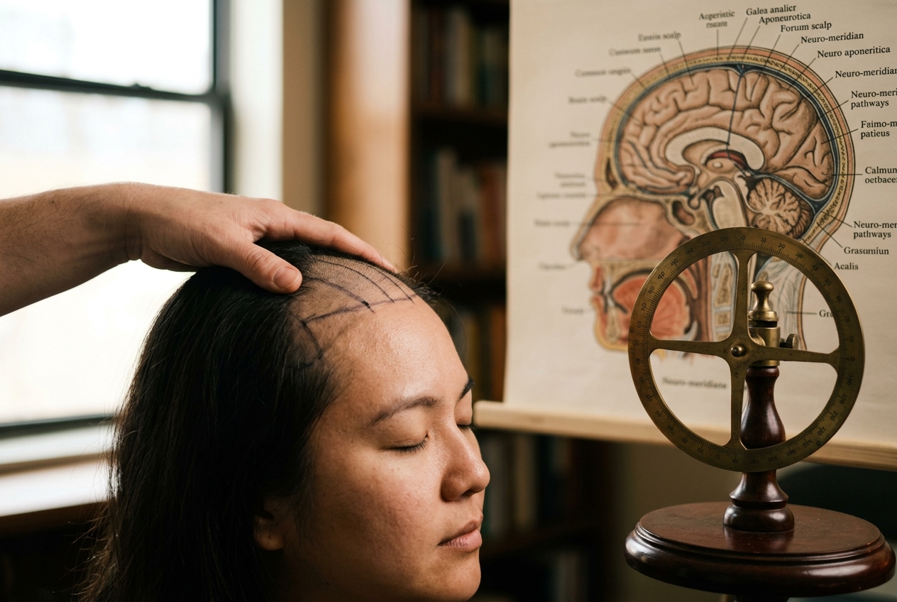
Yet, mapped directly across the convex dome of the human skull lies an extraordinarily responsive bio-electrical switchboard. In the early 1970s, Dr. Jiao Shunfa—a pioneering neurosurgeon practicing in Shanxi Province, China—achieved a profound synthesis that revolutionized contemporary rehabilitative medicine. Rather than viewing the scalp as merely an inert protective sheath of skin, aponeurosis, and periosteum, Jiao recognized that the scalp is a highly sensitive neuro-reflexive projection zone. By correlating classical Chinese medical meridian systems—specifically the Du Mai (Governing Vessel), the Foot Taiyang (Bladder), and the Foot Shaoyang (Gallbladder) channels—with modern neuroanatomical cytoarchitectonics, Dr. Jiao established what is known internationally as Jiao Shunfa Scalp Acupuncture (and its manual analog, linear cranial acupressure).
This system provides a practical, non-invasive anatomical topography for working directly with central nervous system dysfunction. By applying targeted linear mechanosensory pressure across specific cranial projection zones, clinicians and informed individuals can engage the brain’s endogenous capacity for neuroplastic remodeling, upregulate regional cerebral blood flow, and stimulate damaged corticospinal pathways.
2. The Meridian Path — A Journey Through the Cranial Topography
To comprehend the therapeutic potency of Jiao’s scalp cartography, one must explore the anatomical architecture of the cranium, where ancient energetic conduits intersect with the precise somatotopic organization of the human cerebral cortex.
The Primary Surface Coordinate System
Dr. Jiao discarded arbitrary point selection in favor of an absolute cranial coordinate system anchored by two primary anatomical baselines, standardized under the World Health Organization Standard Acupuncture Nomenclature:
- The Anterior-Posterior Sagittal Midline (前后正中线): Traces the midline sagittal suture of the skull, connecting the midpoint between the eyebrows (Yintang / Glabella) directly over the cranial vault to the inferior border of the external occipital protuberance (Inion). This line directly overlies the superior sagittal sinus and mirrors the pathway of the classical Du Mai (Governing Vessel), known in traditional theory as the "Sea of Yang Meridians."
- The Eyebrow-Occipital Baseline (眉枕线): A lateral diagnostic line drawn along the side of the head, connecting the midpoint of the superior border of the eyebrow to the apex of the external occipital protuberance. This line provides the horizontal reference plane against which all functional cortical projection vectors are calibrated.
Classical Channels and Cortical Localization
Classical channel theory posits that the head is where all Yang meridians converge. The Du Mai ascends through the spine, enters the braincase at Fengfu (Du 16), crowns the vertex at Baihui (Du 20), and descends along the forehead at Shenting (Du 24). Flanking this central axis are the bilateral lines of the Foot Taiyang Bladder Channel, which ascend from the inner canthus of the eye, traverse the frontal and parietal bones, and penetrate the occipital cortex. Encircling the temporal parietes is the Foot Shaoyang Gallbladder Channel, weaving across the motor, premotor, and auditory processing regions.
Dr. Jiao recognized that these meridian trajectories correspond to the functional motor and sensory homunculi described by Wilder Penfield in modern neurophysiology (see Penfield's Cortical Homunculus). The cranial zones are not isolated energetic points; they are linear functional projections:
+-----------------------------+-------------------------------------------------------+-------------------------------------------------------------+
| JIAO SHUNFA CRANIAL ZONE    | ANATOMICAL LANDMARKS & TOPOGRAPHY                     | NEUROLOGICAL & CORTICAL CORRELATE                           |
+-----------------------------+-------------------------------------------------------+-------------------------------------------------------------+
| Motor Area (运动区)         | 0.5 cm posterior to AP Midline midpoint to the        | Precentral gyrus; Brodmann Area 4; primary corticospinal    |
|                             | intersection of Eyebrow-Occipital line at temple      | voluntary motor output strip                                |
|                             |                                                       |                                                             |
| - Upper 1/5 Segment         | Superior 1/5 of the Motor Line                        | Lower limb, pelvis, and trunk motor representation          |
| - Middle 2/5 Segment        | Intermediate 2/5 of the Motor Line                    | Upper extremity, hand, wrist, and finger motor control      |
| - Lower 2/5 Segment         | Inferior 2/5 of the Motor Line (Speech Area I)        | Facial muscles, tongue, pharynx, larynx, Broca's area       |
|                             |                                                       |                                                             |
| Sensory Area (感觉区)       | Parallel line located exactly 1.5 cm posterior to    | Postcentral gyrus; Brodmann Areas 1, 2, 3; somatosensory    |
|                             | the Motor Area Line                                   | input processing and tactile proprioception                 |
|                             |                                                       |                                                             |
| Chorea-Tremor Control Area  | Parallel line located exactly 1.5 cm anterior to      | Premotor cortex; Brodmann Area 6; basal ganglia regulatory  |
| (舞蹈震颤控制区)            | the Motor Area Line                                   | circuits for involuntary movement modulation                |
|                             |                                                       |                                                             |
| Vasomotor Area (血管舒缩区) | Parallel line located exactly 1.5 cm anterior to      | Prefrontal vasomotor regulation; cortical control over      |
|                             | Chorea-Tremor Area (3.0 cm anterior to Motor Area)   | peripheral autonomic tone and cerebral perfusion            |
|                             |                                                       |                                                             |
| Vertigo-Auditory Area       | Horizontal line 4 cm long, located 1.5 cm directly    | Superior temporal gyrus; Brodmann Areas 41, 42, 22;         |
| (晕听区)                    | superior to the apex of the auricle (ear tip)         | vestibular nuclei connections and primary auditory cortex   |
|                             |                                                       |                                                             |
| Speech Area II (语言二区)   | Vertical line 3 cm descending from parietal tuber,    | Angular gyrus / Wernicke's area; nominal and sensory        |
|                             | 2 cm posterior-inferior to parietal eminence          | aphasia resolution                                          |
|                             |                                                       |                                                             |
| Speech Area III (语言三区)  | Horizontal line 4 cm extending posteriorly from the   | Posterior temporal-parietal junction; receptive auditory    |
|                             | midpoint of the Vertigo-Auditory Area Line            | language comprehension and semantic decoding                |
|                             |                                                       |                                                             |
| Praxis (Usage) Area (运用区)| Originates at parietal tuber; bifurcates into three   | Supramarginal gyrus; complex motor planning, apraxia        |
|                             | 3 cm rays at 40-degree angles to each other           | correction, and ideational purposeful action                |
|                             |                                                       |                                                             |
| Foot Motor-Sensory Area     | Bilateral lines 1 cm lateral to AP Sagittal Midline,  | Paracentral lobule; medial hemisphere projection of foot,   |
| (足运感区)                  | 3 cm length extending posteriorly from midline offset | lower limb motor-sensory, and bladder/sphincter control     |
+-----------------------------+-------------------------------------------------------+-------------------------------------------------------------+
3. Neurobiological Mechanotransduction & 3 Key Acupressure Zones
The Biological Mechanism: From Scalp Deformation to Synaptic Remodeling
How does mechanical pressure applied to the external scalp induce functional alterations within the cerebral parenchyma shielded behind the dense calvarium? The answer lies in the tri-fold continuum of pericranial fascial mechanotransduction, hemodynamic neurovascular coupling, and trans-cranial neural reflex loops:
- Pericranial Fascial Mechanotransduction: The scalp is composed of five distinct layers (Skin, Dense Connective Tissue, Galea Aponeurotica, Loose Areolar Tissue, and Pericranium). The galea aponeurotica is a tensile, fibroelastic matrix embedded with high densities of low-threshold mechanoreceptors (Ruffini endings, Pacinian corpuscles, and Merkel disks). Mechanical deformation via high-frequency rhythmic linear compression activates mechanically gated ion channels (Piezo1 and Piezo2) within fibroblasts and vascular endothelial cells, triggering downstream intracellular calcium influx and nitric oxide (NO) synthesis (see Mechanisms of Acupuncture and Mechanotransduction).
- Regional Cerebral Blood Flow (rCBF) Upregulation: Trans-cranial vascular communications exist between the superficial temporal, occipital, and supraorbital arteries and the intracranial meningeal and dural sinuses via diploic and emissary veins. Linear acupressure produces localized neurogenic vasodilation through the release of Calcitonin Gene-Related Peptide (CGRP) and Substance P from the trigeminal and cervical cutaneous nerve branches. This peripheral vasodilation induces a reflex autoregulatory upregulation of rCBF within the corresponding underlying ischemic penumbra of the cerebral cortex.
- Neuroplastic Corticospinal Remodeling: Rhythmic sensory inputs directed into specific somatic dermatomes on the scalp propagate along the ascending sensory pathways, projecting through thalamocortical loops back into the primary motor strip (precentral gyrus). In patients recovering from stroke, persistent linear stimulation promotes unmasking of latent horizontal intracortical connections and collateral sprouting across intact fibers of the descending corticospinal tract, fostering functional re-mapping of damaged neural networks.
+---------------------------------------------------------------------------------------------------+
| CLINICAL PROOF: HEMODYNAMIC & NEUROPLASTIC COUPLING                                               |
| Scalp mechanosensory stimulation along Jiao Shunfa lines induces a statistically significant       |
| increase in mean blood flow velocity (Vm) of the middle cerebral artery (MCA) and anterior        |
| cerebral artery (ACA) as measured by Functional Transcranial Doppler (fTCD) and functional MRI.   |
| (Source: Cortical Mechanisms of Scalp Acupuncture, PubMed ID: 24734183)                           |
+---------------------------------------------------------------------------------------------------+
Three Essential Therapeutic Lines for Daily Clinical Practice
Line 1: The Motor Area Line — Contralateral Neuromuscular Reset
- Topographical Location: Locate the midpoint of the Anterior-Posterior Sagittal Midline (halfway between the glabella and inion). Move 0.5 cm posteriorly along the midline to find the upper origin point of the Motor Line. The line terminates antero-inferiorly at the intersection of the Eyebrow-Occipital baseline and the anterior border of the temple hairline (overlying the temporal line of the frontal bone).
- Somatic Palpation: When tracing this oblique vector with the distal pad of the thumb, you will encounter a distinct linear indentation along the galea aponeurotica. Upon arriving at the target segment, firm static palpation will reveal a dull, aching tenderness, often described as a deep, spreading muscular tension or mild sub-galeal warmth.
- Biomechanical Technique: Utilize the radial edge of the thumb or the flexed proximal interphalangeal joint of the index finger. Apply a directional vector gliding force at a 45-degree angle to the calvarium, executing rapid, rhythmic linear oscillations along the specific calibrated segment at a frequency of 100–120 compressions per minute for 2 to 3 minutes:
- Upper 1/5 Segment: Target for lower extremity spasticity, foot drop, and gait instability.
- Middle 2/5 Segment: Target for frozen shoulder, loss of fine motor hand dexterity, and post-stroke wrist drop.
- Lower 2/5 Segment: Target for facial nerve paresis, masticatory spasm, and expressive motor speech blockages.
- Indications: Essential for contralateral hemiparetic motor deficits, chronic postural muscle weakness, occupational repetitive strain, and executive psychomotor fatigue.
Line 2: The Vertigo-Auditory Area — Vestibular Equilibrium & Sensory Processing
- Topographical Location: Measure precisely 1.5 cm directly superior to the apex of the auricle (the superior tip of the folded external ear). Mark a horizontal line 4 cm in total length, extending 2 cm anteriorly and 2 cm posteriorly from this central point, running parallel to the Eyebrow-Occipital baseline.
- Somatic Palpation: Palpating the temporoparietal fascia directly above the ear reveals a distinct vascular texture (superficial temporal vessels) and an acute, bruised tenderness when the vestibular or auditory apparatus is hyper-sensitized or fatigued.
- Biomechanical Technique: Position the index and middle fingertips flat against the 4 cm horizontal line. Apply moderate, firm inward pressure toward the squamosal suture of the temporal bone, followed by linear horizontal friction gliding back and forth across the 4 cm span. Maintain an oscillation rate of 100–120 compressions per minute for 2 minutes bilaterally.
- Indications: Relieves acute positional vertigo, Meniere’s-type disequilibrium, tension-induced tinnitus, vestibular migraine, and sensory overload caused by auditory overstimulation.
Line 3: The Foot Motor-Sensory Area — Bilateral Central Axis Stabilization
- Topographical Location: Located on the crown of the head. Identify the midpoint of the Anterior-Posterior Sagittal Midline. Trace 1 cm anterior to this midpoint, then move 1 cm laterally to the left and right of the sagittal midline. From these bilateral starting points, draw two 3 cm lines extending directly posteriorly, running strictly parallel to the sagittal midline.
- Somatic Palpation: This region overlies the sagittal suture and paracentral lobule. Firm palpation creates a unique sensation of structural decompression across the vertex, frequently eliciting a tingling proprioceptive response that radiates down the spinal column into the calves and plantar surfaces of the feet.
- Biomechanical Technique: Place the thumbs simultaneously on the left and right Foot Motor-Sensory lines. Apply firm downward compressive force while executing simultaneous bilateral linear gliding strokes from anterior to posterior along the 3 cm tracks. Maintain continuous, non-slipping fascial contact with an oscillating rhythm of 100 compressions per minute for 3 minutes.
- Indications: Critical for bilateral lower extremity paresis, lumbar spine instability, neurogenic urinary urgency or frequency, pelvic floor weakness, and loss of somatic grounding during acute panic.
4. A Targeted Protocol: Post-Stroke Hemiparesis and Expressive Speech Rehabilitation
+---------------------------------------------------------------------------------------------------+
| CLINICAL INTERVENTION BLUEPRINT: CEREBROVASCULAR REHABILITATION                                    |
| Objective: Upregulate contralateral corticospinal motor drive and activate peri-Sylvian speech    |
| networks in patients exhibiting upper/lower limb hemiparesis and motor dysphasia.                  |
+---------------------------------------------------------------------------------------------------+
When managing complex neurological deficits such as post-ischemic hemiparesis complicated by expressive speech dysfunction, single-point stimulation is clinically insufficient. A sequential, multi-line synergistic protocol must be executed to recruit collateral neuroplastic pathways.
Complete Clinical Procedure
- Patient Positioning: The subject should be seated comfortably in a supported, high-backed chair with cervical support, or positioned semi-recumbent at a 45-degree angle. Ensure all restrictive neckwear is loosened to optimize jugular venous drainage and carotid arterial perfusion.
- Phase 1: Galeal Warming & Pericranial Mobilization (1 Minute):
- Place both palms flat against the lateral aspects of the cranium over the temporoparietal zones.
- Apply gentle, rhythmic circular mobilization to the scalp relative to the underlying skull bone. This reduces basal pericranial muscular tension, decompresses the superficial veins, and prepares the galea aponeurotica for mechanotransductive signaling.
- Phase 2: Contralateral Motor Area Activation (3 Minutes):
- Target: Locate the Motor Area line contralateral (opposite) to the affected hemiplegic side.
- Execution: Focus predominantly on the Upper 1/5 (for the lower limb) and the Middle 2/5 (for the arm and hand). Using the flexed knuckle of the index finger, deliver firm, unyielding linear gliding strokes from the superior-posterior origin downward and forward along the line. Maintain a cadence of 100–120 compressions per minute. Maintain consistent pressure to compress the subcutaneous tissue against the calvarium without causing skin abrasions.
- Phase 3: Speech Circuit Recruitment (3 Minutes):
- Target: The Lower 2/5 of the Motor Line (Speech Area I) on the dominant hemisphere (typically the left hemisphere for right-handed individuals), followed by Speech Area II (a 3 cm vertical line descending from the parietal tuber).
- Execution: Apply sustained linear friction along Speech Area I, focusing on the release of fascial adhesions near the temporal line. Transition immediately to Speech Area II, utilizing the thumb pad to perform downward vertical linear friction. Instruct the patient to simultaneously attempt vocalization or word repetition to enhance task-specific neuroplastic recruitment.
- Phase 4: Bilateral Foot Motor-Sensory Integration (2 Minutes):
- Target: The paired 3 cm lines situated 1 cm lateral to the sagittal midline at the vertex.
- Execution: Engage both lines simultaneously using dual-thumb glide vectors from anterior to posterior. This recruits bilateral motor-sensory representation of the lower limbs and stabilizes axial postural tone.
- Phase 5: Centering and Grounding (1 Minute):
- Conclude by applying sustained, stationary thumb pressure directly to Baihui (Du 20) at the central vertex. Hold this point with firm, downward pressure while instructing the patient to take four slow, diaphragmatic breaths, visualizing motor intent streaming downward through the brainstem into the affected peripheral extremities.
5. Safety, Neuro-Physiological Boundaries & Clinical Sense
Absolute and Relative Contraindications
- Acute Intracranial Hemorrhage: In the hyper-acute phase of hemorrhagic stroke (within the first 48 to 72 hours), vigorous scalp stimulation is contraindicated until intracranial pressure has stabilized and active re-bleeding has been radiographically ruled out via CT/MRI.
- Structural Calvarial Defects: Never apply compressive force over unhealed craniotomy sites, skull fractures, open cranial wounds, or fontanelles in infants.
- Severe Scalp Infection or Ulceration: Avoid direct stimulation across areas of active cellulitis, folliculitis, or uncharacterized cranial skin lesions.
- Pregnancy Considerations: While scalp acupressure does not engage the aggressive uterine-contractile reflexes associated with lower-body points such as Sanyinjiao (SP 6) or Hegu (LI 4), strong downward mechanical vectors along the Du Mai sagittal midline and vertex (Baihui) should be applied with gentle, balancing pressure rather than heavy, sedating force in individuals with a history of recurrent miscarriage.
+---------------------------------------------------------------------------------------------------+
| SOMATOSENSORY CALIBRATION: THERAPEUTIC "DE QI" VS. TISSUE DAMAGE                                  |
| • CORRECT "DE QI" SENSATION: A profound, radiating ache; localized sensation of warmth;          |
|   sub-galeal pulsation; tingling traveling along peripheral motor pathways.                       |
| • INCORRECT / HARMFUL SENSATION: Sharp, superficial skin tearing; agonizing periosteal pinching;   |
|   dizziness or vasovagal syncope resulting from excessive mechanical force.                       |
+---------------------------------------------------------------------------------------------------+
When to Seek Professional Intervention
Self-applied or caregiver-administered linear acupressure serves as a powerful adjunctive therapy, but it is not a substitute for comprehensive neurological care. Immediate emergency medical consultation is required if a patient presents with sudden-onset facial drooping, unilateral arm weakness, slurred speech (the standard FAST protocol), thunderclap headache, or acute loss of consciousness. For long-term neuro-rehabilitation, this manual protocol should ideally be integrated alongside clinical scalp acupuncture administered by a licensed neuro-acupuncturist, speech-language pathologist, and physical therapist.
6. Today's Takeaway: The Two-Minute Executive Reset
You do not need an acute clinical diagnosis to benefit from Dr. Jiao’s neuroanatomical insights. If you are reading this while experiencing mental exhaustion, tension-induced brain fog, or cognitive overwhelm at your desk, execute this rapid Two-Minute Executive Reset right now:
By bridging the centuries-old wisdom of the Governing Vessel with the precise functional topography of the precentral gyrus, Jiao Shunfa scalp acupressure transforms our understanding of the cranium from a passive bony container into an active, accessible interface for human neurological resilience and recovery.
Authoritative References & Scientific Foundations
- World Health Organization: Standard International Acupuncture Nomenclature
- National Center for Biotechnology Information (NCBI): Neurobiological Mechanisms of Acupuncture
- PubMed: Cortical Mechanisms and Hemodynamics of Scalp Acupuncture
- Wikipedia: Penfield's Cortical Homunculus and Functional Localization
- Wikipedia: Brodmann Cytoarchitectonic Areas of the Cerebral Cortex
- Wikipedia: Corticospinal Tract Anatomy and Physiology