Electroacupuncture Therapy: Navigating Frequency-Specific Neuromodulation, Endogenous Opioid Pathways, and Precision Acupressure Protocols
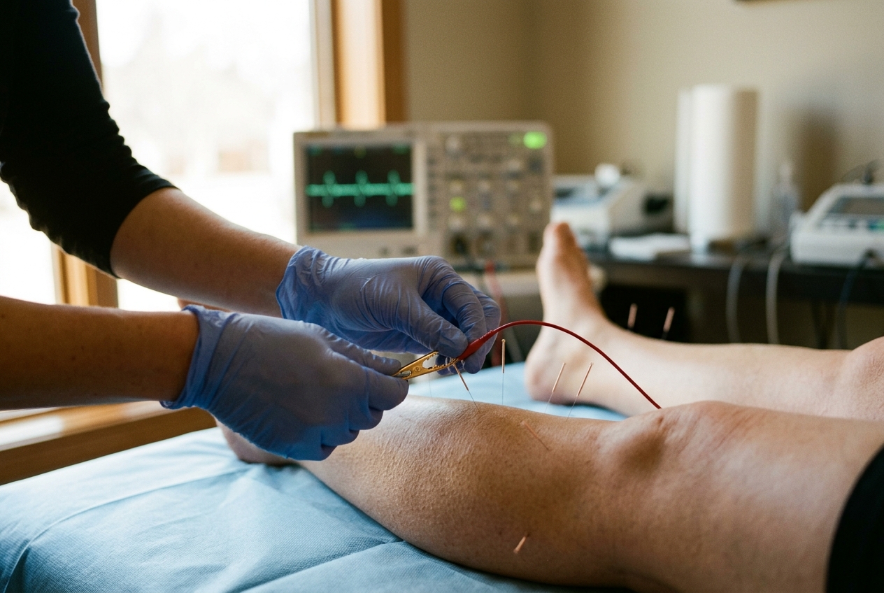
If you sit at a desk in the late afternoon and feel an intractable tightening across the nape of your neck, a hollow visceral distension following ordinary meals, or a refractory state of sympathetic hypervigilance that precludes restorative sleep, you are experiencing the somatic toll of autonomic dysregulation. Modern clinical neurology increasingly conceptualizes these ubiquitous syndromes not as isolated organ pathologies, but as systemic breakdowns in homeostatic feedback loops. Chronic psychosocial stressors and sedentary posturing trigger a persistent upregulation of the hypothalamic-pituitary-adrenal (HPA) axis, tonic sympathetic outflow, and localized neurogenic inflammation within dense fascial compartments.
For millennia, Traditional Chinese Medicine (TCM) systematized these visceral-somatic reflexes along an interconnected lattice of channels or jingluo. Today, contemporary neurophysiology and bioelectronic medicine have converged upon these ancient pathways, revealing that peripheral mechanical and electrical stimulation along classical meridian corridors engages definitive neuroanatomical, neurochemical, and biophysical axes.
By applying targeted microcurrents, dense-disperse electrical oscillations, or structured transcutaneous pressure to specific neurovascular hubs, we do not merely manipulate subjective sensation; we drive precise neuropeptidergic cascades in the spinal cord, recruit somatic motor units, and restore autonomic equilibrium.
BIOELECTRIC CARTOGRAPHY: Fascial Cleavage Planes and Interstitial Low-Impedance Conduits
Far from being esoteric constructs, classical acupuncture meridians map with striking fidelity along intermuscular connective tissue planes, peripheral nerve trunks, and neurovascular bundles. Landmark anatomical dissections and ultrasonic tissue characterization demonstrate that more than 80 percent of classical acupoints coincide with the interfascial cleavage planes of loose connective tissue rich in interstitial fluid, collagen bundles, and unmyelinated nerve terminals.
From a biophysical standpoint, these longitudinal channels serve as preferential electrical conduits within the biological matrix. Biological tissues are anisotropic; current flows preferentially along lines of lowest resistance. Research documented in NCBI PubMed Central studies on acupuncture mechanisms demonstrates that meridian pathways exhibit significantly lower electrical impedance and higher skin capacitance compared to surrounding non-channel cutaneous zones. This low-impedance phenomenon is largely governed by the hydraulic conductivity of interstitial fluid networks:
- Extracellular Matrix Composition: The interstitial channels are rich in glycosaminoglycans and hyaluronic acid, whose fixed negative charges organize water into liquid-crystal hydration shells that facilitate rapid, long-range proton and ion transport (the Grotthuss mechanism).
- Piezoelectric Mechanotransduction: Collagen fibers within the fascial sheath possess intrinsic piezoelectric properties. Mechanical deformation (via manual needle rotation or firm acupressure) generates localized electrical streaming potentials that depolarize adjacent mechanoreceptors and low-threshold afferent fibers.
- Purinergic Signaling Cascades: Physical or electrical distortion of fibroblasts activates stretch-activated ion channels (such as PIEZO1 and PIEZO2) and integrin-cytoskeletal linkages. This triggers a localized efflux of adenosine triphosphate (ATP), which is rapidly cleaved by ectonucleotidases into adenosine. As demonstrated by research on adenosine A1 receptor mechanotransduction, adenosine acts directly upon local A1 receptors on sensory nerve terminals to suppress nociceptive signaling before ascending spinal transmission can occur.
When navigating these pathways on the physical body, one need not rely strictly on classical proportional measurement units (cun). Instead, somatic palpation reveals these channels as natural anatomical depressions, muscular borders, and neurofascial grooves:
- The Leg-Yangming (Stomach) Pathway: Commences beneath the orbit, traverses the anterior border of the masseter muscle, descends over the anterior clavicular space, continues through the belly of the rectus abdominis, passes down the anterolateral thigh, and runs down the lateral crest of the shinbone—specifically within the palpable groove between the tibialis anterior muscle and the extensor digitorum longus—before terminating at the lateral second toe.
- The Leg-Taiyin (Spleen) Pathway: Originates at the medial border of the great toe, tracks along the medial longitudinal arch of the foot, ascends immediately behind the posterior border of the tibia (the inner shinbone, where fatigue and fluid stagnation accumulate after sustained standing), passes the medial aspect of the knee, and ascends along the anteromedial thigh into the abdomen.
- The Arm-Yangming (Large Intestine) Pathway: Begins at the radial index finger tip, crosses the dorsal web space between the first and second metacarpal bones, ascends the lateral aspect of the forearm along the groove of the brachioradialis muscle, traverses the lateral epicondyle of the humerus, ascends the anterior border of the deltoid, and terminates adjacent to the contralateral nasal ala.
- The Arm-Jueyin (Pericardium) Pathway: Arises in the chest, passes down the midline of the anterior arm, traverses the center of the cubital fossa, descends directly down the middle of the inner forearm between two prominent wrist flexor tendons, enters the center of the palm, and exits at the tip of the middle finger.
- The Leg-Jueyin (Liver) Pathway: Originates at the lateral nail bed of the great toe, traverses the dorsal foot within the depression between the first and second metatarsal bones, ascends anterior to the medial malleolus, crosses behind the Spleen meridian along the inner leg, and courses through the medial femoral groove into the hypochondriac region.
MECHANISTIC FOUNDATIONS: Ji-Sheng Han's Frequency Specificity & Peptidergic Neurobiology
The transformation of acupuncture from an empirical discipline into an exact neurobiological science was catalyzed by the pioneering research of Professor Ji-Sheng Han at Peking University. Han demonstrated that the analgesic and physiological actions of electroacupuncture (EA) are mediated by the central nervous system through frequency-dependent synthesis and release of specific endogenous opioid neuropeptides, as detailed in Han's landmark review in Trends in Neurosciences.
Low-Frequency Stimulation (2 Hz) and Central Mu/Delta Agonism
When electro-stimulation is delivered at a low pulse frequency (typically 2 Hz with a pulse width of 0.2–0.5 ms), peripheral myelinated $A\beta$ and $A\delta$ afferent fibers transmit rhythmic, non-noxious action potentials into the dorsal horn of the spinal cord. This input projects rostrally through the spinothalamic tract and parabrachial nucleus to the periaqueductal gray (PAG) and the arcuate nucleus of the hypothalamus.
In response, the central nervous system mobilizes $\beta$-endorphin from pro-opiomelanocortin (POMC) neurons and [Met]-enkephalin within interneuronal pools. These endogenous ligands bind with high affinity to $\mu$ (mu) and $\delta$ (delta) opioid receptors. The resulting activation triggers potent descending inhibitory pathways that travel via the rostral ventromedial medulla (RVM) and the dorsolateral funiculus back to the spinal dorsal horn, releasing serotonin (5-HT) and norepinephrine to suppress ascending nociceptive transmission. This system produces a broad, generalized, long-lasting systemic analgesia and marked autonomic calming.
High-Frequency Stimulation (100 Hz) and Spinal Kappa Agonism
Conversely, delivering continuous high-frequency stimulation at 100 Hz shifts the neurochemical equilibrium entirely. High-frequency pulses do not engage the hypothalamic $\beta$-endorphin cascade; instead, they selectively trigger the release of dynorphins within the deep laminae of the spinal dorsal horn.
Dynorphin acts selectively upon $\kappa$ (kappa) opioid receptors located on the presynaptic terminals of primary sensory afferents. Kappa-opioid activation closes voltage-gated N-type calcium channels and opens inwardly rectifying potassium channels, blocking the presynaptic exocytosis of excitatory neurotransmitters—chiefly Substance P, calcitonin gene-related peptide (CGRP), and glutamate. High-frequency 100 Hz stimulation thus provides rapid, highly potent segmental analgesia, making it particularly effective for acute localized musculoskeletal spasms and visceral inflammatory pain.
The Dense-Disperse (2/100 Hz) Paradigm and Tolerance Prevention
A critical clinical limitation of continuous single-frequency stimulation is the rapid development of neurochemical tolerance: continuous 2 Hz or 100 Hz electroacupuncture over 30 to 45 minutes leads to opioid receptor desensitization and endocytosis.
Han resolved this limitation by designing the dense-disperse (alternating) waveform, in which a 2 Hz train alternates automatically with a 100 Hz train every 3 seconds. This rhythmic cycling induces simultaneous, continuous release of all three endogenous opioid families—$\beta$-endorphin, enkephalins, and dynorphins—saturating $\mu$, $\delta$, and $\kappa$ receptors simultaneously. The alternation delays central receptor downregulation, generating a synergistic, enduring therapeutic effect far superior to either isolated frequency.
Motor Point Recruitment and Neuromuscular Resetting
Beyond central peptidergic mechanisms, electro-stimulation directly targets peripheral neuromuscular physiology. Classical acupoints situated over muscle bellies (such as ST36 over the tibialis anterior or LI4 over the first dorsal interosseous) correspond directly to anatomical motor entry points—the discrete zones where motor nerve branches enter the epimysium.
Applying an electrical potential across these sites depolarizes low-threshold alpha motor neurons, eliciting involuntary, rhythmic, synchronous muscle contractions. This mechanical twitching: * Clears stagnant metabolic byproducts (lactic acid, bradykinin) by driving localized microvascular perfusion; * Mechanically disrupts persistent actin-myosin cross-bridges within myofascial trigger points; * Recalibrates muscle spindle sensitivity and Golgi tendon organ feedback, relieving chronic protective muscular splinting.
Modality Spectrum: Invasive EA, TEAS, and Rhythmic Manual Acupressure
The biophysical principles governing invasive needle electroacupuncture translate directly across a spectrum of modalities:
- Invasive Needle Electroacupuncture (EA): Utilizes filiform stainless-steel needles inserted directly into interfascial boundaries or motor points, delivering precise microcurrents with negligible impedance from the stratum corneum. It remains the gold standard for maximal spatial precision and deep motor-nerve recruitment.
- Transcutaneous Electrical Acupoint Stimulation (TEAS): Employs surface hydrogel electrodes placed over identical acupoints, delivering identical biphasic waveforms (e.g., 2/100 Hz dense-disperse). While the stratum corneum presents higher capacitive resistance (requiring higher voltage amplitudes and clean skin preparation), clinical trials indexed in the World Health Organization Guidelines on Traditional Medicine demonstrate that TEAS achieves comparable endogenous opioid release and autonomic modulation non-invasively.
- Rhythmic Manual Acupressure: Simulates low-frequency (1.5–2 Hz) electrical oscillations through rhythmic, oscillatory mechanical compression (approximately 90 to 120 cycles per minute). This periodic tissue deformation drives cyclic piezoelectric streaming potentials and continuous adenosine A1 receptor agonism, offering a widely accessible, needle-free therapeutic tool.
FIVE ESSENTIAL SOMATIC ACCELERATORS: Neuroanatomy, Deqi Sensations, and Transcutaneous Protocols
The clinical efficacy of acupoint stimulation relies on obtaining the characteristic Deqi sensation—a complex sensory feedback phenomenon indicating successful depolarization of deep $A\delta$ (Group III) and unmyelinated C (Group IV) afferent fibers, accompanied by mechanical "needle grasp" from local collagen remodeling. Below is an academic appraisal of five primary loci, complete with landmark-guided localization and non-invasive application protocols.
1. ST36 (Zusanli — "Leg Three Miles")
- Anatomical Localization: Located on the anterolateral aspect of the lower leg. To locate without measurement rods: sit with the knee flexed at 90 degrees; locate the prominent lower edge of the kneecap (patella); place the index finger of the opposite hand in the soft depression just outside the patellar ligament (the lateral "eye" of the knee). Measure four horizontal finger-widths downward from this depression, and one finger-breadth laterally off the sharp anterior crest of the shinbone (tibia). The point lies directly in the fleshy belly of the tibialis anterior muscle.
- Neuroanatomical Correlate: Deep peroneal nerve, branches of the tibial recurrent artery/vein, motor point of the tibialis anterior. Recent seminal neurobiological work shows that ST36 uniquely activates the vagal-adrenal anti-inflammatory axis, triggering systemic dopamine release from chromaffin cells to curb hyper-inflammatory cytokine storms.
- Characteristic Deqi Sensations: A profound, dull, distending ache accompanied by a warm, heavy sensation that radiates downward along the lateral lower leg into the dorsum of the foot.
- Clinical Applications: Postprandial gastric bloating, chronic functional fatigue, generalized inflammatory down-regulation, and peripheral lower-limb motor rehabilitation.
- Transcutaneous Protocol: Apply sustained, firm perpendicular thumb pressure while executing micro-rotational kneading at a consistent 2 Hz rhythm (two circular pulses per second). Maintain for 2 to 3 minutes per limb. When using a home TEAS device, set to 2/100 Hz dense-disperse mode for 20 minutes at an intensity that produces a visible, comfortable muscle twitch of the tibialis anterior.
2. LI4 (Hegu — "Joining Valleys")
- Anatomical Localization: Situated on the dorsum of the hand. Bring the thumb and index finger flush together; a small muscular prominence rises at the base of the webbing. The point lies at the highest summit of this muscular bulge. Alternatively, open the hand relaxed; place the first interphalangeal crease of the opposing thumb onto the web margin between the thumb and index finger, and pivot the thumb tip down onto the radial aspect of the second metacarpal bone: the point lies directly at the midpoint of that bone's radial shaft.
- Neuroanatomical Correlate: Superficial radial nerve, motor branch of the first dorsal interosseous muscle, deep branch of the ulnar nerve. Mechanistically linked via trigeminal-spinal nuclear projections to descending facial and cephalic analgesia.
- Characteristic Deqi Sensations: An intense, spreading, sour ache, often accompanied by an electrical tingling sensation that courses into the palm and up the radial forearm toward the elbow.
- Clinical Applications: Tension-type and cervicogenic headaches, temporomandibular joint (TMJ) dysfunction, frontal sinus pressure, and generalized systemic pain management.
- Transcutaneous Protocol: Grip the patient's or your own hand with the thumb on the dorsal point and the index finger supporting the palmar surface. Direct the force diagonally inward toward the second metacarpal bone rather than straight through the soft webbing. Exert firm, steady ischemic compression for 90 seconds, followed by 60 seconds of low-frequency circular friction.
3. PC6 (Neiguan — "Inner Gate")
- Anatomical Localization: Positioned on the anterior (volar) aspect of the forearm. Turn the palm upward and make a loose fist while flexing the wrist slightly backward to reveal two prominent, cord-like tendons running down the center of the wrist: the flexor carpi radialis (lateral) and the palmaris longus (medial). Place three fingers of the opposing hand across the inner wrist crease; the point lies directly proximal to the index finger, squarely centered in the groove between these two tendons.
- Neuroanatomical Correlate: Median nerve trunk, anterior interosseous nerve, palmar cutaneous branch. Projects directly to the nucleus of the solitary tract (NTS) in the medulla oblongata, regulating dorsal vagal efferent outflow to the heart and gastrointestinal tract.
- Characteristic Deqi Sensations: A distinct, spreading numbness, light pressure-ache, and subtle electric paresthesia that propagates down into the third digit or ascends the anterior arm to the cubital crease.
- Clinical Applications: Acute nausea, motion sickness, psychogenic palpitation, nocturnal anxiety, insomnia, and functional epigastric discomfort.
- Transcutaneous Protocol: Use the tip of the thumb to hook deeply between the two flexor tendons in a direct perpendicular vector. Apply continuous, calm, non-oscillating ischemic pressure for 2 full minutes while taking slow, deep diaphragmatic breaths (4-second inhalation, 7-second hold, 8-second exhalation) to amplify parasympathetic vagal entrainment.
4. SP6 (Sanyinjiao — "Three Yin Intersection")
- Anatomical Localization: Located on the medial aspect of the lower leg. Find the highest prominence of the inner ankle bone (medial malleolus). Lay four fingers horizontally across the inner calf, starting with the pinky finger resting on the malleolar apex. The point sits directly above the index finger, in a tender muscular groove located immediately behind the posterior border of the shinbone (tibia).
- Neuroanatomical Correlate: Tibial nerve trunk, saphenous nerve, posterior tibial artery and veins; confluence of the Spleen, Liver, and Kidney meridian pathways. Connects directly with pelvic parasympathetic splanchnic nerves (S2–S4).
- Characteristic Deqi Sensations: A pronounced, deeply resonant tenderness, accompanied by a heavy aching sensation that travels up the medial border of the calf and into the pelvic floor.
- Clinical Applications: Menstrual cramping (dysmenorrhea), pelvic venous congestion, lower-extremity fluid retention, nocturnal restlessness, and insomnia.
- Transcutaneous Protocol: Press the thumb or index finger deeply behind the posterior edge of the tibia, angling slightly forward against the bone rather than backward into the calf muscle belly. Apply rhythmic, wave-like compression (3 seconds on, 1 second off) for 2 to 3 minutes per leg.
5. LV3 (Taichong — "Great Surge")
- Anatomical Localization: Located on the top (dorsum) of the foot. Place your finger in the soft webbing between the big toe and the second toe. Slide your finger upward along the groove between the two long foot bones (the first and second metatarsals) toward the ankle for approximately two finger-widths, until your finger drops into a distinct, highly tender V-shaped junction where the bones meet.
- Neuroanatomical Correlate: Deep fibular (peroneal) nerve, dorsal venous arch, first dorsal metatarsal artery. Exerts strong sympatholytic actions, downregulating central vascular resistance and mitigating sympathetic hyperarousal.
- Characteristic Deqi Sensations: A sharp, highly focal, dispersing ache that frequently elicits a rapid local twitch response and radiates across the metatarsal arch.
- Clinical Applications: Cephalic throbbing, temporal migraine, eye strain, irritability, emotional stress, and elevated vascular tension.
- Transcutaneous Protocol: Use the thumb tip to press firmly into the bony angle between the metatarsals. Perform an assertive, downward-gliding stroke toward the toe web, repeating this unidirectional vector 30 to 40 times over 2 minutes, maintaining steady downward force to disperse vascular and muscular tension.
COMPREHENSIVE POINT CHARACTERISTICS & CLINICAL SUMMARY
CLINICAL PROTOCOLS & TRANSLATIONAL INTEGRATION
To translate these individual neurobiological hubs into systemic clinical results, practitioners utilize strategic point combinations that engage complementary ascending and descending neural circuits.
Protocol: The "Neuro-Somatic Reset" for Occipito-Cervical Strain and High-Stress Hypervigilance
When long hours of computational work generate a combination of occipital headache, trapped cervical muscular tension, and an agitated mental state, apply the classical "Four Gates" (Siguan) protocol, augmented by autonomic regulators:
- Preparation and Posture: Recline in a semi-recumbent position with the cervical spine supported by an ergonomic roll, arms resting comfortably at your sides, knees slightly bent over a bolster. Eliminate harsh overhead light.
- Phase 1 — The Distal Four Gates (Minutes 0:00–4:00):
- Begin with LI4 (Hegu) bilaterally. Apply firm, steady compression angled toward the index metacarpal for 90 seconds per hand to trigger descending trigeminal inhibition.
- Transition immediately to LV3 (Taichong) on both feet. Use slow, downward-gliding thumb strokes for 90 seconds per foot. The combination of LI4 (stimulating upper-extremity motor afferents) and LV3 (stimulating lower-extremity deep fibular afferents) produces systemic sympatholysis and vascular relaxation.
- Phase 2 — Autonomic Vagal Stabilization (Minutes 4:00–6:00):
- Engage PC6 (Neiguan) on the left arm. Apply constant, perpendicular thumb pressure for 2 minutes while pacing your respiration to six cycles per minute (a 4-second nasal inhale followed by a 6-second soft mouth exhale). This entrains heart rate variability (HRV) and amplifies vagal cardiac control via median nerve-NTS projections.
- Phase 3 — Long-Loop Descending Analgesia (Minutes 6:00–10:00):
- Conclude by stimulating ST36 (Zusanli) bilaterally. Knead the tibialis anterior muscle belly with steady 2 Hz circular oscillations for 2 minutes per leg. This sustains central $\beta$-endorphin release, consolidates somatic relaxation, and downregulates visceral inflammatory tone.
Transcutaneous Electrical Device (TEAS) Parameters for Clinical Settings
For practitioners and patients utilizing dual-channel electro-stimulation or TENS/TEAS devices: * Electrode Placement: Place Channel 1 hydrogel pads across bilateral LI4 and ST36; place Channel 2 hydrogel pads across bilateral PC6 and SP6. * Waveform Parameters: Select an asymmetric biphasic square wave to prevent electrochemical polarization under the skin. * Frequency Modulation: Program a 2/100 Hz dense-disperse wave (3 seconds at 2 Hz alternating with 3 seconds at 100 Hz). * Pulse Width: Set between 0.2 ms and 0.3 ms. * Intensity Titration: Gradually increase output amplitude until the patient reports a robust, comfortable, non-painful pulsing sensation with visible, subtle rhythmic fasciculations of the target muscles. * Duration: 20 to 30 minutes per clinical session.
SAFETY, CONTRAINDICATIONS, AND CLINICAL JUDGMENT
While non-invasive acupressure and low-voltage surface stimulation have exceptional safety profiles, rigorous clinical practice requires strict adherence to physiological contraindications and red-flag boundaries:
+------------------------------------------------------------------------------------+
|                         ABSOLUTE & RELATIVE CONTRAINDICATIONS                      |
+====================================================================================+
| 1. IMPLANTED CARDIAC DEVICES:                                                      |
|    Electrical stimulation (EA/TEAS) is strictly contraindicated across the torso,   |
|    cervical region, or upper extremities in patients with cardiac pacemakers,      |
|    implantable cardioverter-defibrillators (ICDs), or deep brain stimulators       |
|    due to risks of electromagnetic interference and current leakage.               |
+------------------------------------------------------------------------------------+
| 2. PREGNANCY RESTRICTIONS:                                                         |
|    Points that vigorously promote descending pelvic outflow and uterine            |
|    contractility—specifically SP6 (Sanyinjiao), LI4 (Hegu), BL60 (Kunlun),         |
|    and BL67 (Zhiyin)—are STRICTLY CONTRAINDICATED during pregnancy, except during   |
|    active labor under qualified obstetrical supervision.                           |
+------------------------------------------------------------------------------------+
| 3. LOCAL TISSUE INTEGRITY:                                                         |
|    Never apply electrical current or firm ischemic compression over areas of       |
|    active cutaneous ulceration, infected tissue, acute phlebitic thrombosis,       |
|    unconsolidated bone fractures, or localized oncological neoplasms.               |
+------------------------------------------------------------------------------------+
| 4. SENSORY NEUROPATHIES:                                                           |
|    In patients with advanced diabetic peripheral neuropathy or syringomyelia,      |
|    attenuated thermal and nociceptive feedback requires conservative electrical    |
|    amplitude titration to prevent electrothermal skin irritation.                  |
+------------------------------------------------------------------------------------+
Recognizing Pressure Thresholds: Ischemia vs. Therapeutic Deqi
A common clinical mistake in manual acupressure is applying excessive, bruising force. Therapeutic acupoint stimulation requires mechanical depth and focus, not brute force. * Optimal Pressure: Elicits a distinct, heavy, deep sourness (Deqi) that feels productive and manageable, allowing the patient's breathing to remain slow and uninhibited. * Excessive Pressure: Triggers sharp, superficial, defensive pain that causes reflex muscle bracing, breath-holding, or localized tissue bruising. Such defensive reactions stimulate the sympathetic "fight-or-flight" response, counteracting the intended parasympathetic and analgesic benefits.
When to Seek Specialist Care
Self-administered transcutaneous acupressure and home TEAS are effective adjuncts for functional tension, postprandial dyspepsia, and mild sleep disturbances. However, patients presenting with "red flag" symptoms—such as sudden-onset "thunderclap" headaches, progressive unilateral neurological deficits, unexplained visceral weight loss, severe chest pain radiating to the jaw or arm, or refractory joint swelling—require immediate diagnostic evaluation by a licensed medical physician rather than self-care.
TODAY'S TAKEAWAY: The 120-Second Parasympathetic Interruption
You can directly engage your nervous system's endogenous calming pathways right now, before stepping away from this article.
+------------------------------------------------------------------------------------+
|                    THE 120-SECOND PC6 PARASYMPATHETIC INTERRUPT                    |
+------------------------------------------------------------------------------------+
| 1. LOCATE: Place three fingers across your inner left wrist crease. Find the spot   |
|    just above your index finger, between the two firm middle wrist tendons.         |
|                                                                                    |
| 2. ENGAGE: Press your right thumb firmly inward until you feel a deep, heavy,      |
|    spreading ache (the median nerve Deqi response).                                 |
|                                                                                    |
| 3. BREATHE: Maintain steady, unmoving pressure for 120 seconds while performing     |
|    4-7-8 diaphragmatic breathing (Inhale 4s -> Hold 7s -> Exhale 8s).             |
|                                                                                    |
| 4. PHYSIOLOGICAL RESULT: Engages the nucleus tractus solitarius, resets autonomic  |
|    tone, and downregulates acute sympathetic stress within two minutes.            |
+------------------------------------------------------------------------------------+
By systematically combining the anatomical precision of classical meridian pathways with the biophysical rigor of modern neurobiology, electroacupuncture and transcutaneous point stimulation bridge the gap between ancient clinical intuition and contemporary neuroscience. Whether delivered via invasive filiform needles, modern biphasic microcurrents, or simple manual compression, targeted stimulation along these fascial conduits remains one of medicine's most potent tools for restoring physiological equilibrium.
Authoritative Scientific References & Further Reading
- National Institutes of Health (NIH) Consensus Conference Statement on Acupuncture — Exhaustive clinical review of acupuncture efficacy, neurobiological validity, and safety profiles.
- Han, J.S. (2003). Acupuncture: Neuropeptide release produced by electrical stimulation of different frequencies. Trends in Neurosciences — The definitive paper establishing 2 Hz ($\beta$-endorphin/enkephalin) versus 100 Hz (dynorphin) frequency specificity.
- Goldman, N., et al. (2010). Adenosine A1 receptors mediate local anti-nociceptive effects of acupuncture. Nature Neuroscience — Landmark discovery of purinergic signaling, local ATP efflux, and adenosine A1-mediated mechanotransduction during somatic point rotation.
- World Health Organization (WHO) Traditional, Complementary and Integrative Medicine Technical Documents — Global clinical guidelines, point standardization, and safety protocols for acupuncture-related modalities.
- National Center for Biotechnology Information (NCBI) Review: Neurobiological Foundations of Acupuncture and Electroacupuncture — Comprehensive academic analysis of low-impedance interstitial fascial conduits, descending RVM/PAG inhibition, and autonomic balance.
- Wikipedia Academic Compendium: Electroacupuncture — Detailed historical overview, frequency parameters, and clinical translation of modern electroceutical therapies.