Bloodletting Techniques (Ci Luo Fang Xue Fa) Meridian Dynamics: Governing Superficial Micro-Venipuncture, Toxic Heat Purging, Microvascular Stasis Resolution, and Hemodynamic Decompression in Traditional Chinese Medicine
Abstract & Pedagogical Objective
This curriculum chapter provides a comprehensive, academically rigorous exploration of Cì Luò Fàng Xuè Fǎ (刺络放血法, Collateral Pricking and Bloodletting Therapy), one of the oldest surgical-energetic modalities within Traditional Chinese Medicine (TCM). Rooted in the foundational treatises of the Huángdì Nèijīng (黄帝内经), collateral bloodletting serves to decompress stagnant microvascular beds (Tōng Luò 通络), eliminate blood stasis (Qū Yū 祛瘀), drain toxic heat (Xiè Rè Jiě Dú 泄热解毒), and re-establish systemic homeostatic balance.
By integrating classical channel theory with modern vascular biology, hemorheology, and neuroimmunology, this chapter details the biomechanical instrumentation, modern physiological cascades, anatomical loci protocols, and aseptic safety parameters essential for safe, evidence-based clinical application as recognized within global frameworks by the World Health Organization (WHO) Traditional Medicine Strategy.
1. Classical Foundations & Theoretical Origin of Collateral Pricking
The therapeutic evocation of micro-hemorrhage occupies a central position in classical Chinese medicine. Far from being a crude or non-specific historical practice, collateral bloodletting represents a targeted hemodynamic intervention designed to alter channel pressure gradients and restore the laminar flow of Qì (气) and Xuè (Blood, 血).
1.1 The Canonical Mandate of the Huángdì Nèijīng
The theoretical underpinnings of Cì Luò are established throughout the Huángdì Nèijīng (Yellow Emperor's Inner Classic, c. 1st–2nd Century BCE).
- The Nine Classical Needles (Jiǔ Zhēn 九针): In Líng Shū (灵枢, Divine Pivot) Chapter 1 (Jiǔ Zhēn Shí'èr Yuán 九针十二原) and Chapter 10 (Jīng Mài 经脉), the classical needle spectrum is cataloged. Among them, the Lance or Three-Edged Needle (Fēng Zhēn 锋针) is designated explicitly for collateral evacuation:
"The sixth is called the Feng Zhen (lance needle); it has a triangular body with sharp edges, designed to discharge chronic diseases and drain superficial blood collaterals."
- The Principle of Eliminating Stasis: Sù Wèn (素问, Basic Questions) Chapter 62 (Diào Jīng Lùn 调经论 — Treatise on Regulating the Meridians) articulates the primary therapeutic axiom:
“血实者,决之” (Xuè shí zhě, jué zhī — "Where there is blood repletion, drain it with rapid evacuation.")
- Clearing Ancient Stagnation: Líng Shū Chapter 5 (Gēn Jié 根结) further corroborates:
“宛陈则除之” (Wǎn chén zé chú zhī — "That which is stagnant, accumulated, and old must be eliminated.")
In classical physiology, the circulatory matrix consists of the deep, longitudinal Primary Channels (Jīng Mài 经脉) and the superficial, horizontally arborizing Collateral Network (Luò Mài 络脉). When pathogenic factors (such as Wind-Heat, Toxic Fire, or Phlegm) penetrate the exterior, or when chronic emotional constraint impairs Qì propulsion, the Luò Mài become engorged. The blood, unable to circulate freely, congeals into dark, static blood (Yū Xuè 瘀血), giving rise to localized pain, tissue distension, intractable heat, or neurological disturbance.
1.2 Energetic Mechanics: Tong Luo and Toxic Heat Dispersion
The physiological action of Cì Luò Fàng Xuè Fǎ operates through three distinct meridian mechanisms:
- Unblocking Stagnant Collaterals (Tōng Luò 通络): By mechanically perforating the distended superficial venules (Luò Cì 络刺), localized high-resistance resistance zones are bypassed. The sudden drop in peripheral vascular resistance re-establishes normal pressure differentials between the Jīng Mài and Luò Mài, allowing healthy Qì and nutritive Yíng (营) fluids to reperfuse ischemic micro-domains.
- Purging Pathogenic Toxic Heat (Xiè Rè Jiě Dú 泄热解毒): Toxic heat (Rè Dú 热毒) possesses an ascending, hyper-dynamic quality that agitates the Xuè layer. Pricking terminal branch collaterals provides an immediate physiological and energetic outlet, venting pathological heat outward through the skin rather than permitting it to sink inward into the Zàng-Fǔ (脏腑) organs or pericardium (Xīn Bāo 心包).
- Harmonizing Yin-Yang and Dynamic Polarity: In acute emergency states—such as stroke, syncope, or febrile convulsions—excess Yang flares uncontrollably to the cranium while Yin collapses. Pricking the terminal Jing-Well (Jǐng 井) points rapidly vents pathogenic Yang repletion, restoring functional equilibrium across the central nervous axis.
2. Instrumentation & Modality Classification
The application of Cì Luò is modulated by specific instrumentation, penetration kinetics, and clinical objectives. Three primary procedural modalities dominate the clinical landscape.
| Needle Modality | Classical Nomenclature | Mechanical Design | Primary Puncture Technique | Clinical Objective |
|---|---|---|---|---|
| Three-Edged Needle | Sān Léng Zhēn (三棱针) | Rigid stainless steel shaft with a tri-pyramidal bevel and three cutting edges | Spot Pricking (Diǎn Cì Fǎ 点刺法) / Clustered Pricking (Cù Cì Fǎ 促刺法) | Rapid micro-venipuncture of engorged superficial venules; targeted bloodletting (1–5 drops). |
| Cutaneous / Plum Blossom Needle | Méi Huā Zhēn (梅花针) / Qī Xīng Zhēn (七星针) | Flexible hammer containing a circular cluster of 5 to 7 micro-fine surgical pins | Scattered / Tapping Pricking (Sǎn Cì Fǎ 散刺法 / Kòu Cì Fǎ 叩刺法) | Broad, superficial dermal micro-trauma without deep venipuncture; diffuse capillary extravasation. |
| Prick-and-Cup Apparatus | Cì Luò Bá Guàn (刺络拔罐) / Wet Cupping | Combined Three-Edged / Plum Blossom lance with negative-pressure glass or polycarbonate cups | Puncture followed by Vacuum Extravasation | Deep regional stasis mobilization; extraction of 2–15 mL of interstitial and microvascular exudate. |
2.1 The Three-Edged Needle (Sān Léng Zhēn) and Spot Pricking (Diǎn Cì Fǎ)
The modern Sān Léng Zhēn is the direct descendant of the classical Fēng Zhēn. Engineered from high-tensile surgical steel, its triangular apex produces a small tri-radiate or slit incisional puncture that remains patent long enough to permit capillary decompression before spontaneous platelet plug formation occurs.
- Spot Pricking (Diǎn Cì Fǎ): The primary technique for discrete acupoints (e.g., Jing-Well points, Ěrjiān EX-HN6, Tàiyáng EX-HN5). The operator stabilizes the skin, executes a microsecond dart-like thrust (depth: 1.0–3.0 mm), and immediately retracts the needle.
- Clustered / Aggregated Pricking (Cù Cì Fǎ): Multiple spot pricks applied circumferentially around a focal pathological lesion (such as a local carbuncle, localized neurodermatitis plaque, or stubborn hematoma) to relieve compartmental tension.
2.2 Cutaneous / Plum Blossom Needling (Méi Huā Zhēn)
The cutaneous needle leverages an elastic handle to deliver perpendicular, rapid-fire strikes across wider dermatomal zones. By flicking the wrist, the operator creates hundreds of minute, uniform epidermal perforations without penetrating the subcutaneous vascular plexuses. This modality disperses exterior Wind-Heat, stimulates hair follicles in alopecia areata, and regulates autonomic dysregulation along the Paravertebral Huatuojiaji (Huátúo Jiājǐ 华佗夹脊) loci.
2.3 Prick-and-Cup Therapy (Cì Luò Bá Guàn Fǎ)
Documented globally across historic medical systems—including the Greco-Arabic Hijama tradition reviewed extensively on PubMed / NCBI—wet cupping combines cutaneous puncture with negative atmospheric pressure (typically −150 to −300 mmHg). The applied suction draws static venous blood, inflammatory cytokines, and interstitial fluid out of the deep muscular layers toward the surface, making it effective for chronic myofascial syndromes and thoracic-lumbar blood stasis.
3. Modern Physiological & Hemorheological Mechanisms
Contemporary clinical research has clarified the biological events triggered by collateral micro-venipuncture. Far from functioning solely as an empirical or placebo modality, bloodletting induces rapid hemorheological, vascular, and neuroimmunological shifts.
3.1 Microvascular Decompression and Compartment Kinetics
In tissue zones characterized by chronic inflammation or acute traumatic spasm, the microcirculation exhibits severe capillary hypertension, micro-thrombi formation, and endothelial swelling. This results in local interstitial hypertension and tissue hypoxia.
Micro-puncture of engorged terminal venules: 1. Reduces Microvascular Pressure: Relieves the post-capillary resistance barrier, instantaneously reducing backward venular pressure. 2. Restores Starling Forces: Normalizes trans-capillary fluid filtration, facilitating the clearance of interstitial edema. 3. Improves Capillary Perfusion: Triggers reactive vasodilation via endothelial Nitric Oxide (NO) synthase upregulation, reoxygenating ischemic tissue domains.
3.2 Hemorheology and Endogenous Fibrinolytic Activation
Studies indexed in biomedical databases such as NCBI PMC demonstrate that targeted bloodletting alters local and systemic hemorheological profiles: * Erythrocyte Aggregation: High shear rates induced by sudden local blood drainage disrupt erythrocyte "rouleaux" formations, reducing whole blood viscosity. * Fibrinolysis Cascade: The controlled micro-injury to the vascular endothelium releases endogenous tissue plasminogen activator (t-PA). t-PA catalyzes the conversion of plasminogen to plasmin, accelerating the lysis of microvascular fibrin deposits and clearing local micro-thrombi.
3.3 Neuroimmunological Modulation: Pro-inflammatory Cytokine Suppression
Targeted micro-bloodletting exhibits measurable anti-inflammatory dynamics. Clinical trials examining bloodletting at Ěrjiān (EX-HN6) and Dàzhuī (GV14) demonstrate significant down-regulation of primary circulating and localized pro-inflammatory cytokines:
$$\text{Downregulation Index} = \downarrow [\text{TNF}-\alpha] + \downarrow [\text{IL}-1\beta] + \downarrow [\text{IL}-6] + \uparrow [\text{IL}-10]$$
By attenuating tumor necrosis factor-alpha ($\text{TNF}-\alpha$) and interleukin-6 ($\text{IL}-6$), the systemic inflammatory cascade is interrupted. This blunts the acute phase response, reduces hepatic C-reactive protein (CRP) synthesis, and recalibrates hypothalamic thermoregulatory set-points during acute febrile conditions.
3.4 Somatosensory Pain Gating and Descending Inhibitory Control
The immediate analgesic response observed following collateral bloodletting is governed by neuroanatomical gating mechanisms: * The Gate Control Mechanism: Rapid needle penetration delivers high-intensity mechanical stimulation to myelinated $\text{A}\delta$ (Group III) afferent fibers. These fast-conducting signals reach the dorsal horn of the spinal cord and activate inhibitory interneurons within the substantia gelatinosa, closing the presynaptic gate against incoming, slow, unmyelinated $\text{C}$-fiber (Group IV) nociceptive inputs. * Endogenous Opioid Surges: Ascending spinothalamic stimulation triggers the periaqueductal gray (PAG) and rostral ventromedial medulla (RVM), stimulating the release of endogenous opioid peptides—namely $\beta$-endorphin, dynorphin, and met-enkephalin—while simultaneously depleting localized Substance P stores at the peripheral site of puncture.
4. Anatomical Topography & Locus-Specific Clinical Protocols
The clinical efficacy of Cì Luò Fàng Xuè Fǎ depends on precise point selection based on classical meridian topography and localized vascular architecture.
                    CRANIOFACIAL & CERVICAL PRICKING SITES

                           [Erjian EX-HN6] (Apex of auricle)
                                  \       /
                                   \     /
         [Taiyang EX-HN5] -------- ( o o ) -------- [Taiyang EX-HN5]
         (Temporal fossa venule)     \ = /          (Temporal fossa venule)
                                      |
                                      |
                                  [Shaoshang LU11] (Radial thumb border)
                                  [Shangyang LI1]  (Radial index border)
                                  [Zhongchong PC9] (Tip of middle finger)
                                  [Yinbai SP1]     (Medial big toe border)
4.1 The Terminal Jing-Well Points (Jǐng Xué 井穴)
Located on the extreme distal extremities (the tips of the fingers and toes), the Jing-Well points represent the dynamic origin sites where Qì transitions from deep interior pathways to the exterior channel interface. Their superficial location makes them prime targets for acute emergency revival and the rapid venting of organ heat.
A. Shàoshāng (少商, LU11)
- Anatomical Locus: Radial aspect of the distal phalanx of the thumb, 0.1 cùn posterior to the corner of the nail bed.
- Regional Neurovascular Profile: Branches of the radial digital artery and vein; terminal branches of the lateral antebrachial cutaneous nerve and the superficial branch of the radial nerve.
- Target Pathology: Severe acute pharyngitis (acute tonsillitis), high fever, laryngeal edema, pediatric convulsions, apoplectic coma.
- Execution Protocol: Stabilize the thumb, milk the micro-vessels toward the distal apex to induce capillary pooling, apply rapid perpendicular puncture with a three-edged needle to a depth of 1.0 mm, and express 1 to 3 drops of blood until the color transitions from dark purple-red to bright crimson.
B. Shāngyáng (商阳, LI1)
- Anatomical Locus: Radial aspect of the index finger, 0.1 cùn posterior to the corner of the nail bed.
- Regional Neurovascular Profile: Dorsal digital artery/vein network; terminal digital branches of the median nerve.
- Target Pathology: High febrile states, acute parotitis (mumps), severe dental pulpitis, acute loss of consciousness.
- Execution Protocol: Rapid spot prick, express 2 to 3 drops of blood; frequently combined with LU11 to clear Yangming and Taiyin channel heat.
C. Zhòngchōng (中冲, PC9)
- Anatomical Locus: In the center of the tip of the middle finger (or 0.1 cùn from the radial nail corner).
- Regional Neurovascular Profile: Palmar digital arteriovenous anastomosis; terminal sensory arborizations of the median nerve.
- Target Pathology: Apoplexy (stroke resuscitation), cardiac heat exhaustion, acute heatstroke, fainting, severe febrile delirium.
- Execution Protocol: Central apex puncture, express 1 to 2 drops of blood.
D. Yǐnbái (隐白, SP1)
- Anatomical Locus: Medial aspect of the great toe, 0.1 cùn posterior to the corner of the nail bed.
- Regional Neurovascular Profile: Medial dorsal digital vessels; superficial peroneal nerve and medial plantar nerve branches.
- Target Pathology: Acute dream-disturbed sleep, mania (Kuáng Zhèng 狂证), chronic non-healing uterine bleeding (via moxibustion), acute abdominal distension, severe pediatric fright.
- Execution Protocol: Spot prick to yield 1 to 2 drops of blood to unbind Spleen constraint and clear disturbed Shen.
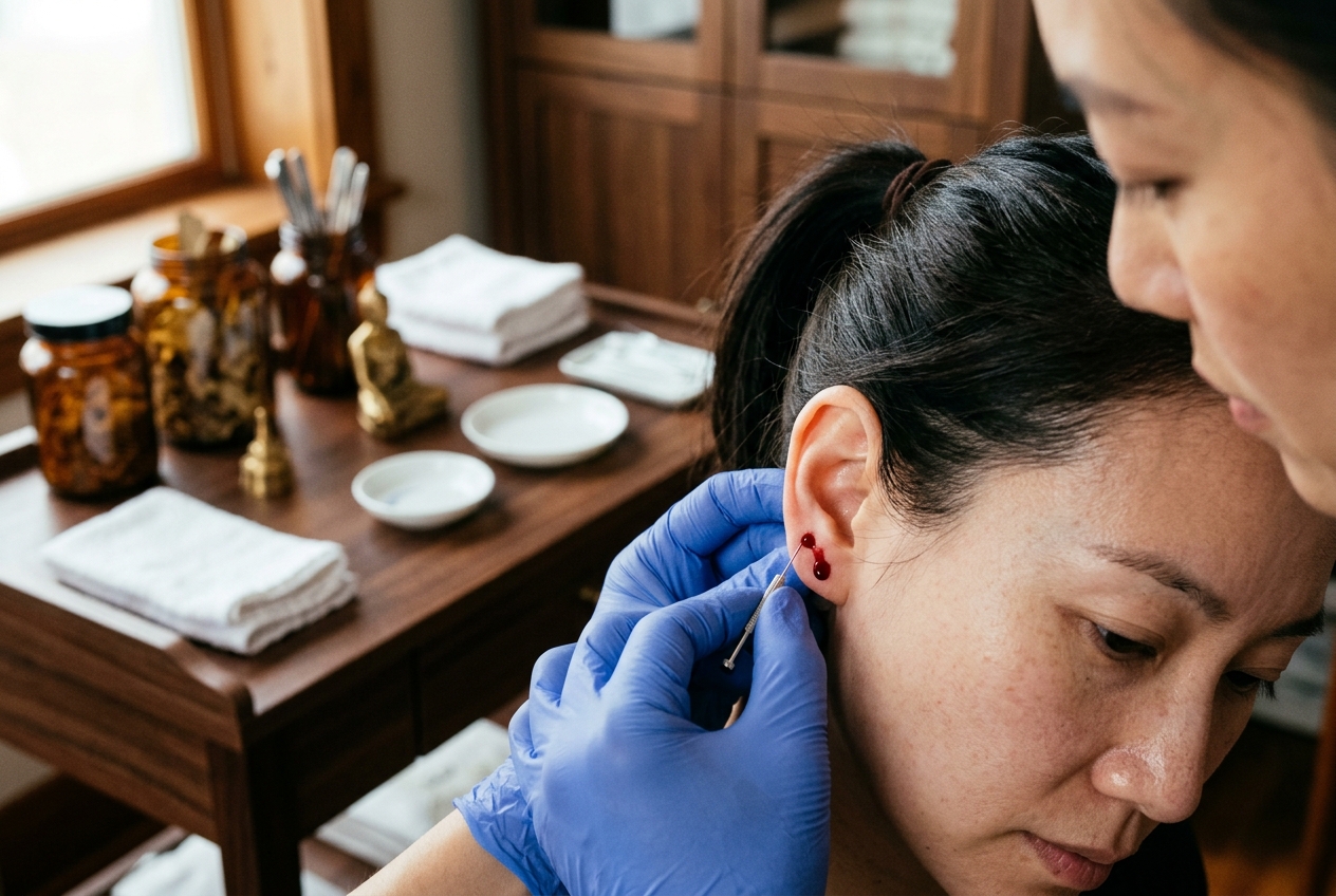
4.2 Ear Apex (Ěrjiān 耳尖, EX-HN6)
- Anatomical Locus: At the apex of the auricle, discovered by folding the ear forward toward the tragus; the point lies at the highest anatomical peak of the helix.
                           EAR APEX (Erjian EX-HN6)
                                   (•) EX-HN6 (Top of Helix when folded)
                                 .---.
                               /       \
                              |   ( )   |  <-- Auricular Cartilage
                              |   / \   |
                               \ '---' /
                                '---'
- Regional Neurovascular Profile: Branches of the superficial temporal artery and posterior auricular artery/vein; branches of the auriculotemporal nerve and great auricular nerve.
- Target Pathology: Acute hypertensive emergencies (used as an adjunct therapy), acute epidemic conjunctivitis (pink eye), acute temporal/parietal migraines, severe facial acne vulgaris, insomnia due to hyperactive Liver Fire.
- Execution Protocol: 1. Rub the entire earlobe and auricle vigorously with sterile gauze until distinct hyperemic flushing is observed. 2. Fold the auricle anteriorly along its vertical midline to isolate the exact apex. 3. Stabilize the skin firmly between the operator's non-dominant thumb and index finger. 4. Perform a swift vertical prick (depth: 1.0–2.0 mm) with a disposable Three-Edged Needle or 21G lancet. 5. Squeeze the adjacent auricular tissue gently from base to apex, expressing 3 to 5 drops of dark blood. Wipe each droplet with a sterile dry cotton sphere.
4.3 Popliteal Fossa Venules (Wěizhōng 委中, BL40)
- Anatomical Locus: Midpoint of the transverse popliteal crease, between the tendons of the biceps femoris and semitendinosus muscles.
- Regional Neurovascular Profile: Superficial subcutaneous venous plexus emptying directly into the small saphenous vein and popliteal vein; posterior femoral cutaneous nerve and tibial nerve trunk deeply situated.
- Target Pathology: Acute lumbar muscle sprain (Yāo Tòng 腰痛), chronic lumbar disc herniation with lower-limb radiculopathy, lower-limb varicosities with severe stasis dermatosis, erysipelas (Dān Dú 丹毒), systemic blood-heat psoriasis and eczema.
- Execution Protocol: 1. Patient is positioned in the prone position with lower extremities relaxed and extended. 2. Apply a venous tourniquet or manual pressure proximal to the popliteal fossa if needed to engorge superficial venules. 3. Inspect the popliteal fossa under focused direct lighting to identify dark, distended, twisted superficial micro-venules (Luò Mài). 4. Disinfect the region broadly with povidone-iodine or 75% isopropyl alcohol. 5. Using a Three-Edged Needle, execute an angled or perpendicular puncture (depth: 1.5–3.0 mm) directly into the lumen of the engorged venule. 6. Permit the dark venous blood to discharge freely into a kidney dish or sterile gauze pad (5 to 10 drops, or up to 2–5 mL if utilizing a secondary vacuum glass cup). 7. When the effluent transitions from dark burgundy to bright arterial-red, compress the venule with sterile gauze for 2–3 minutes to secure hemostasis.
4.4 Tàiyáng (太阳, EX-HN5)
- Anatomical Locus: In the temporal region, in the depression approximately 1.0 cùn posterior to the midpoint between the lateral end of the eyebrow and the outer canthus of the eye.
- Regional Neurovascular Profile: Superficial temporal artery and vein; zygomaticofacial and auriculotemporal nerves; motor branches of the facial nerve.
- Target Pathology: Intractable temporal migraines, cluster headaches, acute ocular hyperemia, optic neuritis, trigeminal neuralgia (ophthalmic/maxillary branch).
- Execution Protocol: Palpate the temporal depression to locate superficial venules. Anchor the surrounding dermis. Execute a precise 1.0–2.0 mm spot prick into the venule or apply prick-and-cup technique. Express 2 to 5 drops of dark blood. Apply firm post-procedural compression.
5. Step-by-Step Clinical Methodology, Hemostasis & Safety Governance
The execution of Cì Luò Fàng Xuè Fǎ requires strict adherence to aseptic protocols, precise tissue mechanics, and thorough pre-procedural contraindication screening.
5.1 Procedural Execution Steps
- Patient Positioning: * Absolute Rule: Patients must never receive bloodletting while standing. * Position the patient in a comfortable supine or prone posture. If seated (e.g., for Ěrjiān or craniofacial points), provide a supportive arm-rest and headrest to eliminate the risk of fall injuries caused by vasovagal syncope (Zhēn Yùn 针晕).
- Local Congestion Maneuver: * Prior to puncturing, use the thumb and forefinger to massage, stroke, or milk the tissue surrounding the target point toward the distal tip. This pools venous blood locally, dilating the micro-collaterals and ensuring an immediate yield upon puncture.
- Aseptic Skin Preparation: * Cleanse the locus with an alcohol swab (75% isopropyl alcohol) or povidone-iodine solution. Apply in concentric outward circles over an area of at least 3 cm diameter. Allow to dry completely (30 seconds) to ensure microbial eradication and prevent stinging.
- Mechanical Insertion Dynamics: * Secure the target site between the non-dominant thumb and forefinger to create skin tension. * Hold the sterile, single-use Three-Edged Needle or 21–25G lancet with the dominant hand in a dart-grip. * Execute a rapid, sub-second perpendicular or slightly oblique thrust to a controlled depth of 1.0 to 3.0 mm. Immediate high-velocity retraction is required to minimize pain perception and prevent laceration of the deeper periosteum or neural sheaths.
- Controlled Droplet Quantification and Expression: * Allow the first droplet to emerge spontaneously. * If flow stops prematurely, apply gentle, rhythmic perifocal pressure starting 1–2 cm away from the puncture site, stroking toward the aperture. * Do not directly pinch, twist, or brutally squeeze the puncture margins, as this causes localized hemolysis, tissue damage, and subcutaneous ecchymosis. * Express the prescribed quantity: 1 to 5 drops for Jing-Well, Ěrjiān, and craniofacial points; up to 1–5 mL for large popliteal or wet-cupping applications.
- Hemostasis and Post-Care: * Immediately upon reaching the desired droplet threshold, apply firm, direct pressure with a sterile, dry cotton ball or gauze compress for 60 to 120 seconds (or longer if clinical parameters require). * Verify complete hemostasis. Apply a sterile adhesive dressing if necessary. Instruct the patient to keep the puncture site clean and dry for at least 12 hours.
5.2 Clinical Safety Governance & Contraindications
Rigorous Contraindication Checklist
Micro-venipuncture and bloodletting therapies are strictly regulated interventions. Practitioners must assess each patient against the following absolute and relative contraindications before treatment.
Absolute Contraindications
- Hemorrhagic Disorders & Coagulopathies: Conditions such as Hemophilia A/B, von Willebrand disease, and disseminated intravascular coagulation (DIC) carry high risks of persistent, non-compressible hemorrhage.
- Severe Thrombocytopenia: Platelet count $< 50 \times 10^9/\text{L}$ ($<50,000/\mu\text{L}$) presents an unacceptably high risk of bleeding complications.
- Active Anticoagulant / Antiplatelet Pharmacotherapy: Patients on therapeutic doses of Warfarin, Direct Oral Anticoagulants (DOACs: Apixaban, Rivaroxaban, Dabigatran), Heparin, or Dual Antiplatelet Therapy (Aspirin + Clopidogrel) must not undergo bloodletting due to impaired primary and secondary hemostasis.
- Severe Anemia: Hemoglobin concentrations $< 8.0\text{ g/dL}$ reflect systemic red blood cell deficiency, making any intentional blood volume depletion counter-therapeutic.
- Pregnancy (High-Risk Zones): Bloodletting is strictly contraindicated on the lower abdomen, lumbosacral regions, and strong down-bearing points (e.g., Hégǔ LI4, Sānyīnjiāo SP6, Jiānjǐng GB21, Zhìyīn BL67) due to the risk of stimulating uterine contractions.
- Local Cutaneous Pathologies: Never puncture directly into active skin ulcers, deep burn eschars, active erysipelas lesions, or areas with superficial vascular malformations (e.g., cavernous hemangiomas or arterial aneurysms).
Relative Precautions & Adverse Event Management
- Vasovagal Syncope (Zhēn Yùn): Manifests as pallor, cold diaphoresis, dizziness, nausea, tachycardia transitioning to bradycardia, and hypotension.
- Immediate Action: Terminate the procedure, recline the patient horizontally into the Trendelenburg position (legs elevated), provide warm fluids, and apply firm nail pressure or moxibustion to Rénzhōng (GV26) and Bǎihuì (GV20).
- Local Hematoma Formation: Results from inadequate post-puncture compression or transection of deeper venules.
- Management: Apply direct firm compression for 5 minutes. Apply a cold compress for the first 24 hours to limit extravasation, followed by warm compresses after 24 hours to promote vascular reabsorption.
6. Comprehensive Summary Table of Clinical Bloodletting Protocols
| Locus Name & Code | Anatomical Position | Target Clinical Indications | Needle Type & Method | Standard Discharge Volume | Key Safety Precaution |
|---|---|---|---|---|---|
| Shàoshāng (LU11) | Radial border of thumb, 0.1 cùn from nail bed | Acute tonsillitis, high fever, laryngeal edema, apoplectic coma | Three-Edged Needle; perpendicular spot prick | 1–3 drops | Pre-massage to pool blood; do not pierce the periosteum. |
| Shāngyáng (LI1) | Radial border of index finger, 0.1 cùn from nail bed | Acute parotitis, high febrile state, dental pulpitis, coma | Three-Edged Needle; perpendicular spot prick | 2–3 drops | Quick dart-like thrust to minimize pain. |
| Zhòngchōng (PC9) | Central apex tip of the middle finger | Acute heatstroke, cardiac syncope, pediatric convulsions, coma | Three-Edged Needle; axial terminal puncture | 1–2 drops | Avoid excessive squeezing; wipe cleanly with sterile cotton. |
| Yǐnbái (SP1) | Medial border of big toe, 0.1 cùn from nail bed | Acute mania, sleep disturbances, acute infantile fright | Three-Edged Needle; spot prick | 1–2 drops | Cleanse thoroughly; feet carry higher bacterial loads. |
| Ěrjiān (EX-HN6) | Apex of the auricular helix when folded | Hypertensive crisis (adjunct), acute conjunctivitis, migraine, acne | Three-Edged Needle / 21G lancet; vertical puncture | 3–5 drops | Vigorously rub the ear first until hyperemic; do not pierce cartilage. |
| Wěizhōng (BL40) | Center of the popliteal crease | Acute lumbar sprain, chronic sciatica, erysipelas, blood-heat eczema | Three-Edged Needle; angled venipuncture $\pm$ cupping | 5–10 drops (or 2–5 mL with cup) | Patient must be prone; do not puncture the deep popliteal artery or tibial nerve. |
| Tàiyáng (EX-HN5) | Temporal fossa, 1.0 cùn posterior to mid-brow/canthus | Temporal migraine, cluster headache, ocular inflammation | Three-Edged Needle; superficial venipuncture | 2–5 drops | Palpate temporal vessels carefully; apply firm 2-minute post-compression. |
7. Curriculum Synthesis & Review
Modern research supports the classical perspective outlined in the Huángdì Nèijīng: Cì Luò Fàng Xuè Fǎ is an efficient and focused hemodynamic intervention. By selecting targeted anatomical loci—from the distal Jing-Well points to the larger venous networks of the popliteal crease—practitioners can relieve localized capillary hypertension, clear microvascular stasis, downregulate systemic inflammatory cytokines, and stimulate descending somatosensory pain inhibition.
Mastery of this modality requires an understanding of both classical channel theory and modern vascular physiology, alongside rigorous attention to aseptic technique, precise needle mechanics, and strict adherence to clinical contraindications.
Authoritative Biomedical References & Further Reading
- World Health Organization (WHO) Traditional Medicine Global Frameworks
- National Center for Biotechnology Information (NCBI) PubMed Central Database
- NCBI PMC Biomedical Literature Repository
- Traditional Chinese Medicine — Clinical and Theoretical Principles (Wikipedia)
- Acupuncture & Collateral Therapeutics Taxonomy (Wikipedia)