Acupotomy Therapy: Navigating Micro-Scalpel Fascial Release, Biomechanical Adhesion Decompression, and Precision Acupressure Protocols
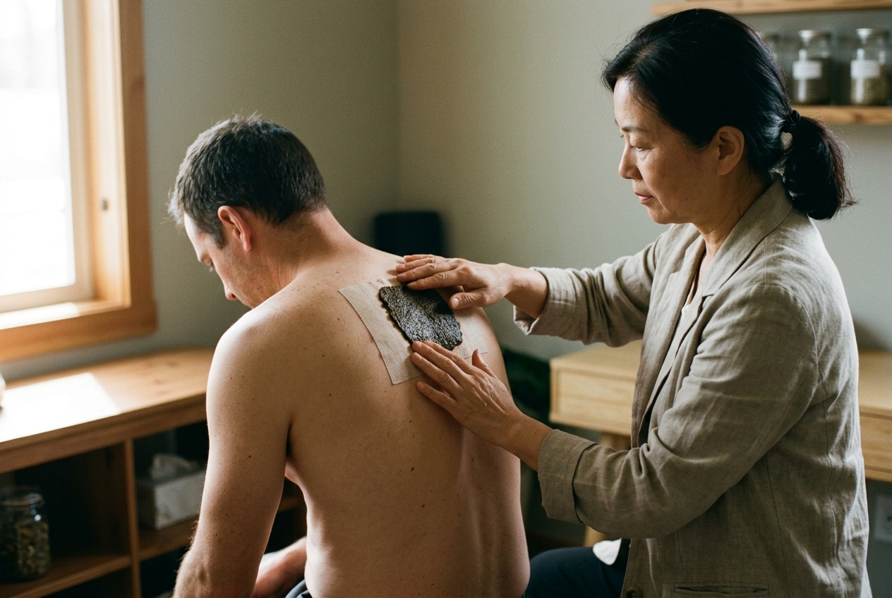
1. Opening — A Problem This Solves Today: The Epidemic of Silent Fascial Adhesion
Consider the ubiquitous physical malady of the twenty-first century: the dull, unyielding ache at the base of the skull, the persistent burning between the scapulae, or the stubborn stiffness of a knee that resists stretching, massage, and pharmacotherapy. Millions of individuals navigate their daily routines under the shadow of chronic myofascial entrapment. Conventional clinical workups frequently present a baffling paradox: standard plain-film radiographs and magnetic resonance imaging (MRI) scans reveal unremarkable or mildly degenerative findings that fail to explain the patient's profound functional limitation.
In conventional conservative care, patients are cycled through nonsteroidal anti-inflammatory drugs (NSAIDs), physical therapy, and standard filiform acupuncture. While these modalities provide transient palliation, the root pathology—structural, mechanical densification and micro-scarring within the deep connective tissue—often remains untouched. Conversely, open surgical decompression remains disproportionately invasive for diffuse soft-tissue contractures.
+-----------------------------------------------------------------------------------+
|                           THE FASCIAL ENTRAPMENT CYCLE                            |
|                                                                                   |
|   Repetitive Strain / Ischemia  -->  Aseptic Exudative Inflammation               |
|              ^                                    |                               |
|              |                                    v                               |
|   Persistent Nociceptive Arc    <--  Fascial Densification & Pathological Cross-Links |
|   & Compartment Hypertension               (Indurated Nodules / "Ashi" Points)    |
+-----------------------------------------------------------------------------------+
This diagnostic and therapeutic void is precisely what acupotomy (Xiao Zhen Dao, literally "small needle-scalpel") is engineered to resolve. Conceived as a minimally invasive bridge between traditional meridian medicine and modern orthopedic surgery, acupotomy directly targets the densified, adhered, and fibrotically altered fascia that encases muscles, tendons, and peripheral nerves. By addressing the physical architecture of chronic soft-tissue damage, this discipline provides a coherent explanatory model and a definitive clinical solution for conditions once dismissed as untreatable muscular strain.
2. Historical Genealogy & Theoretical Synthesis: From the Ling Shu to Zhu Hanzhang
The conceptual lineage of acupotomy represents an extraordinary synthesis of classical Chinese medical antiquity and contemporary surgical anatomy. Far from being a modern rupture from tradition, the needle-knife trace its roots directly to the foundational canon of East Asian medicine: the Huangdi Neijing: Ling Shu (The Yellow Emperor’s Classic of Internal Medicine: Spiritual Pivot, circa 1st century BCE).
Chapter Nine of the Ling Shu delineates the Nine Classical Needles (Jiu Zhen), a sophisticated array of therapeutic instruments engineered for distinct tissue depths and functional objectives. While the fine, flexible Haozhen (filiform needle) became the global face of classical acupuncture, the canon explicitly mandated heavier, bladed instruments for structural pathology:
- The Pi Zhen (Sword/Stiletto Needle): Characterized by a flat, double-edged blade designed to drain purulent exudates, transect stubborn fibrous bands, and excise necrotic tissue.
- The Feng Zhen (Three-Edged Lance Needle): Engineered with triangular cutting edges to lance acute stasis, resolve hematomas, and drain blood stagnations within recalcitrant channels.
For centuries, the use of large, bladed needles declined due to the absence of modern aseptic techniques and anatomical mapping. In 1976, Chinese orthopedic surgeon Professor Zhu Hanzhang revitalized this dormant legacy. Confronted with recalcitrant cases of musculoskeletal contracture and post-traumatic adhesion that resisted both filiform acupuncture and open surgery, Zhu fused the bladed geometry of the ancient Pizhen with the cylindrical shaft of a hypodermic needle.
Zhu established that chronic musculoskeletal disorders are fundamentally mechanical diseases: disorders of micro-adhesion, tissue contracture, scarring, and structural imbalance. By applying a specialized needle with a flat micro-chisel tip measuring between 0.4 mm and 1.2 mm in diameter, practitioners could perform closed micro-surgical lysis, percutaneous tenotomy, and osteofascial decompression without skin incisions, sutures, or general anesthesia.
This innovation earned widespread institutional recognition across East Asia, evolving into a distinct medical sub-specialty documented in authoritative databases including NCBI PubMed and recognized in contemporary complementary frameworks evaluated by the World Health Organization (WHO). Modern research by pioneers like Dr. Helene Langevin on connective tissue mechanics (NCBI PMC Fascia Research) and Dr. Carla Stecco on fascial anatomy has validated Zhu’s foundational hypothesis: fascial planes are dynamic, highly innervated mechanosensory organs that undergo pathological densification under unremitting mechanical stress.
3. Pathophysiology of Chronic Soft Tissue Damage & the Jing Jin (Sinew Channel) Pathways
To understand how acupotomy operates, one must examine the intersection between the classical East Asian paradigm of the Jing Jin (Sinew / Tendino-Muscular Channels) and modern fascial tensegrity.
The Biological Cascade of Fascial Densification
When soft tissue experiences acute trauma or chronic low-grade ergonomic strain, it undergoes an invariant pathophysiological sequence:
- Micro-Trauma & Aseptic Exudation: Microscopic tearing of collagen fibrils within the epimysium, perimysium, or tendon sheaths triggers an aseptic inflammatory cascade. Plasma proteins, fibrinogen, and inflammatory mediators flood the interstitial matrix.
- Pathological Cross-Linking & Hyaluronan Viscosity: In the absence of full functional excursion, fibrin coalesces into dense, disorganized collagenous cross-links. The lubricating hyaluronan layer between fascial layers transforms from a fluid sol state to a viscous, glue-like gel state.
- Fascial Contracture & Nodulation: Fibroblasts differentiate into contractile myofibroblasts, exerting unremitting tension on surrounding extracellular matrix (ECM) scaffolds. This produces palpable indurations, string-like bands (Tiao Suo), and discrete nodules known classically as Jie (knots) or Ju (gatherings), and contemporarily as myofascial trigger points or Ashi loci.
- Ischemic Compression & Micro-Compartment Hypertension: As the fascial sheath thickens and contracts, resting intra-compartmental fluid pressure rises. This pressure compresses thin-walled capillary venules and unmyelinated terminal nerve branches. The result is focal tissue hypoxia, cellular acidosis, accumulation of substance P and calcitonin gene-related peptide (CGRP), and heightened peripheral nociceptor sensitization.
The Jing Jin as Tensegrity Pathways
Classical Chinese medicine mapped these relationships not as isolated muscles, but as unified kinetic highways: the Twelve Sinew Channels. Unlike the primary internal meridians, the Jing Jin originate at the extremities, track along major fascial lines, and "bind" (Jie) at major bony prominences, joint capsules, and aponeuroses. When a single "binding" becomes fibrotic—such as the suboccipital insertion along the Foot Taiyang Sinew Channel or the iliotibial band insertion along the Foot Shaoyang Sinew Channel—the entire global tensegrity chain distorts, radiating abnormal biomechanical vector pulls across the musculoskeletal system.
4. Mechanotransductive Mechanics: Longitudinal Cutting & Transverse Peeling
Acupotomy exerts its clinical effects through direct, micro-mechanical tissue restructuring coupled with downstream neuro-cellular mechanotransduction. Unlike filiform needles, which primarily elicit bioelectrical and neurochemical modulation via minimal mechanical displacement, the needle-knife acts as a precision micro-chisel operating at the exact interface of fascial adhesion.
1. Longitudinal Incisive Releasing (Zong Xing Shu Qie)
The operator aligns the chisel blade strictly parallel to the physiological trajectory of the target muscle fibers, tendon strands, or ligamentous bands. The blade is advanced through the indurated lesion with controlled longitudinal thrusts: * Vector Mechanics: By incising along the longitudinal axis of the contracted tissue, the micro-blade divides transverse pathological cross-links while leaving normal longitudinal structural fibers functionally intact. * Compartment Decompression: This incision breaches the rigid, inextensible fascial envelope, precipitating a drop in interstitial fluid pressure. The mechanical decompression immediately relieves compressive hoop-stress on encased micro-vessels.
2. Transverse Blunt Peeling (Heng Xing Bo Li)
Following longitudinal release, the operator rotates the scalpel blade 90 degrees or employs the blunt shoulder of the instrument to execute gentle lateral sweeping motions across the planar surface: * Interface Liberation: This maneuver physically separates adhered fascial sheets (such as the sliding interface between the deep surface of the trapezius and the underlying splenius capitis). * Shear-Induced Solation: The shearing force breaks up viscous hyaluronan aggregates, transforming the lubricating gel back into an expansive fluid sol, restoring multi-planar gliding excursion.
The Biomolecular & Neurophysiological Cascade
The physical act of needle-knife release initiates a multi-tiered therapeutic cascade:
- Microvascular Reperfusion: Relieving vascular compression instigates an immediate hyperemic flush. Hypoxic tissues receive an influx of oxygenated blood, washing out acidic metabolic byproducts, lactate, and algogenic neuropeptides (Substance P, CGRP, Bradykinin).
- Quenching the Sensitizing Reflex Arc: Chronic fascial tension continuously excites group III and IV mechanosensitive nociceptors, maintaining a state of central spinal cord "wind-up" in the dorsal horn. Transecting the source of continuous tension abolishes this afferent loop, leading to the rapid normalization of neuromuscular tone.
- Fibroblast Mechanotransduction: The localized micro-trauma stimulates resident fibroblasts to upregulate Matrix Metalloproteinases (MMPs), enzymes responsible for degrading aberrant scar tissue and orchestrating the synthesis of well-organized, compliant Type I collagen fibrils over disorganized Type III scar tissue. For deeper technical overviews on these cellular pathways, see literature on NCBI and classical needle dynamics on Wikipedia.
5. Structured Clinical Case Breakdown: Chronic Cervical Spondylopathy & Myofascial Entrapment
To contextualize the practical application of acupotomy, consider this structured clinical case study representative of common cervical pathology.
Diagnostic Palpatory Localization
Pre-procedural examination does not rely solely on imaging; it mandates meticulous, multi-layered static and dynamic palpation along the cervical Sinew Channels: 1. Suboccipital Inserional Enthesopathy: Palpation along the superior nuchal line (territory of GB20 Fengchi and BL10 Tianzhu) detects rock-hard, nodular indurations representing fibrotic contracture of the rectus capitis posterior major and obliquus capitis superior muscle insertions. 2. Levator Scapulae & Upper Trapezius Banding: Transverse strumming over the superior medial angle of the scapula (SI14 Jianwaishu) and the midpoint of the upper trapezius (GB21 Jianjing) reveals distinct, rope-like cords (Tiao Suo) that elicit sharp local tenderness and reproduce the patient's familiar radiating cephalic headache.
Procedural Acupotomy Protocol
- Patient Position: Prone with the cervical spine moderately flexed on a dedicated headrest to expose the suboccipital and cervicothoracic junction.
- Aseptic Preparation: Surgical-grade chlorhexidine antisepsis, sterile fenestrated drape, sterile gloves, and single-use 0.6 mm × 50 mm disposable needle-scalpels.
- Point-by-Point Decompression:
- Site A (Superior Nuchal Line / GB20 locus): The blade is introduced perpendicular to the occipital bone until firm osseous contact is made. The needle is retracted 1 mm into the deep fascial enthesis, where 2–3 precise longitudinal micro-cuts are executed parallel to the cervical axis, followed by delicate lateral peeling across the periosteal attachment.
- Site B (Medial Scapular Angle / Insertion of Levator Scapulae): The blade is inserted down to the periosteum of the scapula, lysing the calcified myofascial insertion with longitudinal slicing motions to eliminate the anchor point of chronic traction.
- Hemostasis & Post-Needling Maneuvers: Direct sterile compression is applied for 2 minutes per site to prevent hematoma formation, followed by passive full-range cervical flexion-distraction and rotational mobilization.
Documented outcomes for these micro-decompression procedures are extensively detailed in contemporary orthopedic research publications on NCBI PubMed: Acupotomy Clinical Research.
6. The Meridian Path — A Journey Through the Sinew Highways
To maintain the architectural integrity of the human frame post-release, one must understand the expansive pathways along which these fascial tensions travel. In Chinese medicine, these lines are the Jing Jin; in modern biomechanics, they correspond precisely to Thomas Myers' Anatomy Trains myofascial meridians.
The Foot Shaoyang (Gallbladder) Sinew Channel: The Lateral Kinetic Highway
The Foot Shaoyang Sinew Channel represents the primary stabilizer of lateral balance and torsional rotation. * The Pathway in the Living Body: It commences at the fourth toe, traverses the top of the foot, and climbs over the prominent outer ankle bone (lateral malleolus). From there, it ascends the outer lower leg along the muscular belly of the peroneals, binding forcefully at the prominent bony knob just below the outer knee—the head of the fibula. * The Torso & Cranial Ascents: The pathway continues upward along the thick, fibrous iliotibial (IT) band on the outer thigh, crosses the hip joint, and scales the lateral rib cage in a zigzag pattern through the intercostal muscles. Ascending across the shoulder crest and the side of the neck, it encircles the ear and anchors onto the lateral skull and forehead. * Everyday Functional Significance: Whenever you shift your weight laterally, brace against uneven ground, or twist your torso, this entire lateral fascial chain engages. When the fibular anchor point tightens, it transmits torque all the way up the lateral thigh and neck, locking the lateral cervical spine into rigid immobility.
The Foot Yangming (Stomach) Sinew Channel: The Anterior Postural Scaffold
Running along the front of the body, the Foot Yangming Sinew Channel governs forward deceleration and sagittal alignment. * The Pathway in the Living Body: Beginning at the middle toes, it travels over the anterior ankle flexor retinaculum, tracks directly up the front of the shinbone (the anterior compartment), binds below the kneecap at the tibial tuberosity, and climbs the powerful quadriceps femoris. Entering the abdominal wall, it forms the rectus sheath, climbs the chest across the pectorals, and anchors into the jaw and facial musculature. * Everyday Functional Significance: This is the primary chain compressed by prolonged seated postures. Shortening of this anterior highway drags the pelvis forward, flattens the thoracic curve, and forces the cervical spine into a compensatory forward-head posture.
7. Key Precision Acupressure Points for Fascial Maintenance
While invasive acupotomy must be performed by a licensed medical specialist, patients and manual therapists can utilize precision acupressure on specific master points to sustain postoperative fascial compliance, stimulate microcirculation, and prevent the recurrence of pathological cross-links.
1. Gallbladder 34 (GB34 — Yanglingquan / "Yang Mound Spring")
- Category: Hui-Meeting Point for all Sinews/Tendons (Jin Hui); He-Sea Point of the Gallbladder Meridian.
- Anatomical Localization: Locate the bony protrusion on the outer side of your leg just below and in front of the knee joint (the head of the fibula). Slide your finger approximately one finger-width forward and slightly downward into the soft, distinct depression between the peroneus longus and extensor digitorum longus muscles.
- Palpatory Sensation (De Qi): When precisely engaged, you will experience a deep, resonant, spreading ache that often sends a light, pleasant tingling sensation down the outer edge of the shin toward the ankle.
- Application Parameters: Apply firm, perpendicular pressure using the tip of the thumb or reinforced middle finger. Maintain steady ischemic pressure for 90 to 120 seconds while performing microscopic circular friction (1–2 mm oscillations).
- Clinical Utility: Resolves generalized muscular tightness, decreases passive myofascial resistance along the lateral kinetic chain, alleviates hip and knee stiffness, and downregulates systemic neuromuscular irritability.
2. Stomach 36 (ST36 — Zusanli / "Leg Three Miles")
- Category: He-Sea Point of the Stomach Channel; Primary Point for Tonifying Qi, Blood, and Post-Injury Tissue Repair.
- Anatomical Localization: Place the palm of your hand over the center of your kneecap with your fingers pointing downward along your shin. The point lies approximately four finger-widths below the lower border of the patella, exactly one thumb-width lateral to the sharp anterior crest of the tibia, in the fleshy belly of the tibialis anterior muscle.
- Palpatory Sensation: A pronounced, heavy, warming, and distending ache that radiates downward toward the dorsal aspect of the foot.
- Application Parameters: Apply sustained, deep, rhythmic compressions. Press firmly for 5 seconds, release slightly for 2 seconds, and repeat this pumping rhythm for 2 to 3 minutes per limb.
- Clinical Utility: Stimulates systemic microvascular perfusion, accelerates extracellular matrix repair, mitigates chronic fatigue, downregulates systemic inflammatory markers, and relieves anterior shin and knee strain.
3. Bladder 40 (BL40 — Weizhong / "Middle of the Crease")
- Category: He-Sea Point of the Bladder Channel; Command Point of the Lumbar Region and Posterior Kinetic Chain.
- Anatomical Localization: Located at the exact midpoint of the transverse crease on the back of the knee joint (the popliteal fossa), situated between the prominent tendons of the biceps femoris and semitendinosus muscles.
- Palpatory Sensation: A distinct, sharp-yet-relieving ache with a nerve-reflex sensation that gently disperses up the hamstring and down the gastrocnemius.
- Application Parameters: While seated with the knee slightly flexed to relax the popliteal fascia, cup both hands around the knee and press your index or middle fingertips upward and inward into the center of the crease. Hold continuous, moderate pressure for 60 to 90 seconds while slowly flexing and extending the ankle.
- Clinical Utility: Decompresses the posterior superficial myofascial chain, relieves acute and chronic low back spasm, alleviates hamstring contractures, and drains venous stasis from the lower extremity.
8. Point Combination Protocol: The "Evening Desk-Worker Decompression"
Prolonged static desk work induces severe myofascial contracture along the suboccipital, levator, and lateral spinal chains. Perform this integrated, synergistic acupressure routine nightly to restore tissue sliding before sleep.
9. Safety, Sense, and Clinical Contraindications
While self-applied acupressure is exceptionally safe, clinical acupotomy is an invasive surgical procedure that demands absolute anatomical precision and strict adherence to medical safety protocols.
+---------------------------------------------------------------------------------------+
|                               CONTRAINDICATION MATRIX                                 |
+---------------------------------------------------------------------------------------+
| ABSOLUTE CONTRAINDICATIONS FOR INVASIVE ACUPOTOMY:                                     |
| • Active systemic or local cutaneous/articular infections.                            |
| • Coagulopathies, hemophilia, or active high-dose anticoagulant therapy.              |
| • Severe uncontrolled diabetes mellitus (risk of compromised wound healing).          |
| • Severe osteoporosis or local osseous malignancies at the target enthesis.          |
| • Anatomical zones with direct vascular/neural exposure (e.g., femoral triangle).     |
+---------------------------------------------------------------------------------------+
| ACUPRESSURE PRECAUTIONS & PREGNANCY WARNINGS:                                         |
| • Strong downward-draining points (e.g., LI4, SP6, GB21) must be strictly avoided     |
|   during pregnancy due to potential uterine contractility effects.                     |
| • "Healthy Pain" vs. "Damaging Pressure": Acupressure should elicit a dull, heavy,   |
|   distending sensation (*De Qi*). Sharp, lancinating, electric-shock pain indicates   |
|   direct nerve impingement; pressure must be released immediately.                    |
| • When to Seek a Specialist: Persistent unremitting pain, progressive neurological    |
|   deficits (numbness, loss of motor strength), or unyielding joint swelling mandate   |
|   formal evaluation by a licensed orthopedic surgeon or acupotomy physician.          |
+---------------------------------------------------------------------------------------+
10. Today's Takeaway: The 2-Minute Sinew Reset
You do not need an entire clinic to experience the neuro-fascial benefits of meridian mechanics. Before closing this chapter, execute this rapid two-minute intervention to reset your postural tensegrity:
+---------------------------------------------------------------------------------------+
|                              THE 2-MINUTE GB34 RESET                                  |
+---------------------------------------------------------------------------------------+
| 1. IDENTIFY: Reach down to your outer right leg and locate the head of the fibula     |
|    just below the knee. Drop your thumb forward and down into the soft hollow of GB34.|
| 2. ENGAGE: Apply firm, sustained pressure until you feel a deep, satisfying ache.     |
| 3. MOBILIZE: While maintaining steady pressure, slowly rotate your right ankle in a   |
|    wide circle—5 times clockwise, 5 times counter-clockwise. Notice how the muscles  |
|    glide beneath your thumb.                                                          |
| 4. REPEAT: Switch to the left leg and repeat the procedure.                           |
| 5. EVALUATE: Stand up and gently rotate your neck and shoulders. Note the subtle yet   |
|    palpable release of tension across your upper back and lateral frame.              |
+---------------------------------------------------------------------------------------+
Through the lens of modern science, the ancient insights of the Ling Shu and the pioneering micro-surgical innovations of Professor Zhu Hanzhang converge on a single truth: our bodies are interconnected kinetic ecosystems. By learning to release the micro-adhesions that bind our tissues, we unlock the body's innate capacity for fluid, pain-free movement.